CD44 and CXCL9 predicting rejection after LTx
Stacks Image 34054
Based on a fruitful collaboration with the department of Visceral, Transplantation, Thoracic, and Vascular Surgery at the University of Leipzig our paper on „CD44 and CXCL9 serum protein levels predict the risk of clinically significant allograft rejection after liver transplantation“ has been accepted for publication in Liver Transplantation. 

Authors are Nathanael Raschzok, Anja Reutzel-Selke, Rosa Bianca Schmuck, Mehmet Haluk Morgul, Ulrich Gauger, Kukuh Aji Prabowo, Laura-Marie Tannus, Annekatrin Leder, Benjamin Struecker, Sabine Boas-Knoop, Michael Bartels, Sven Jonas, Christian Lojewski, Gero Puhl, Daniel Seehofer, Marcus Bahra, Andreas Pascher, Johann Pratschke, and Igor Maximilian Sauer.

The diagnosis of acute cellular rejection (ACR) after liver transplantation is based on histological analysis of biopsies because non-invasive biomarkers for allograft rejection are not yet established for clinical routines. CD31, CD44, and CXCL9 have previously been described as biomarkers for cross-organ allograft rejection. Here, we assessed the predictive and diagnostic value of these proteins as serum biomarkers for clinically significant ACR in the first six months after liver transplantation in a prospective study. The protein levels were measured in 94 patients immediately prior to transplantation, at postoperative days (POD) 1, 3, 7, and 14, and when biopsies were performed during episodes of biochemical graft dysfunction. Our results suggest that CD44 and CXCL9 may serve as predictive biomarkers to identify liver allograft recipients at risk for clinically significant ACR.
Read More
Cover – march issue of Tissue Engineering
Stacks Image 34059

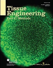
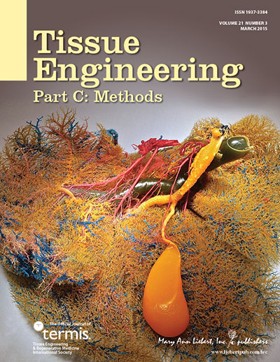
One of the figures of our paper „Porcine liver decellularization under oscillating pressure conditions – A technical refinement to improve the homogeneity of the decellularization process“ made it to the cover of the march issue of Tissue Engineering, Part C : Methods.

Congratulations to Dietrich Polenz, who made the corrosion cast of a decellularized pig liver matrix: red, hepatic artery; blue, portal vein; yellow, bile duct and gallbladder.

Read More
Particles for microRNA-targeted manipulation
Biomaterials accepted our paper on „Micron-sized iron oxide-containing particles for microRNA-targeted manipulation and MRI-based tracking of transplanted cells“. Authors are Annekatrin Leder, Nathanael Raschzok, Christian Schmidt, Duygu Arabacioglu, Antje Butter, Susanne Kolano, Luisa S. de Sousa Lisboa, Wiebke Werner, Dietrich Polenz, Anja Reutzel-Selke, Johann Pratschke, and Igor M. Sauer.

Particle-based delivery systems for therapeutic manipulation and tracking of transplanted cells by magnetic resonance imaging (MRI) are commonly based on nanometer-sized superparamagnetic iron oxide particles (SPIOs). Here, we present a proof of concept for multifunctional, silica based micron-sized iron oxide-containing particles (sMPIO) that combine fluorescence imaging, MRI tracking, and on-the-spot targeting of specific microRNAs on a particle surface for therapeutic manipulation by RNA interference. Antisense locked nucle-LNA) were covalently bound to the surface of silica-based, DAPI-integrated, micron-sized iron oxide particles (sMPIO--LNA). In vitro studies using primary human hepatocytes showed rapid particle uptake (4 hours) that was accompanied by significant depletion of the targeted microRNA Let7g (80%), up- regulation of the target proteins Cyclin D1 and c-Myc, and specific proteome changes. sMPIO--LNA- labeled cells were successfully detected by fluorescence imaging and could be visualized by MRI after intrasplenic transplantation in rats. This new theranostic particle provides a promising tool for cell transplantation where cellular imaging and microRNA-based manipulation is needed.

Biomaterials is the leading journal in its field. Impact factors released by ISI in July 2014 showed Biomaterials with an impact factor of 8.312!
Read More
Presentations at DTG 2014
Stacks Image 34104
Nathanael Raschzok presented his poster „Protein biomarkers for diagnosis and prediction of acute cellular rejection after liver transplantation“, Benjamin Strücker his work entitled „Oszillierende Druckschwankung verbessern signifikant die Ratten- und Schweineleber Dezellularisierung“ and Rosa Schmuck her posters „Risk of postoperative infections after LTX rises with MELD score“ and „Protein biomarkers in bile as a diagnostic tool after liver transplantation“.
Read More
Ben Strücker: Charité Clinical Scientist
Stacks Image 34114
Dr. med Benjamin Strücker successfully applied for the Charité Clinical Scientist 2015 program.
His project is entitled „Humanized Porcine Liver““. Clinical mentor is Prof. Dr. Johann Pratschke, scientific mentors is Priv.-Doz. Dr. med Igor M. Sauer.

The program is supported by Stiftung Charité which was endowed by the entrepreneur Johanna Quandt in order to promote biomedical "knowledge entrepreneurs" that is, change makers in biomedicine at the Charité. The goal of this program is to develop new career paths in clinical specialist medical training. The focus of the training program "Clinical Scientist" is translational research ("bench-to-bedside") which will be realized by a reduction in clinical routine and an improved curriculum with defined goals.
Read More
Sham Laparotomy and microRNA Expression in Rats
Stacks Image 34124
BMC Research Notes accepted our latest paper on „Independent Effects of Sham Laparotomy and Anesthesia on Hepatic microRNA Expression in Rats“ for publication.

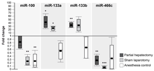
Studies on liver regeneration following partial hepatectomy (PH) have identified several microRNAs (miRNAs) that show a regulated expression pattern. These studies involve major surgery to access the liver, which is known to have intrinsic effects on hepatic gene expression and may also affect miRNA screening results. We performed two-third PH or sham laparotomy (SL) in Wistar rats to investigate the effect of both procedures on miRNA expression in liver tissue and corresponding plasma samples by microarray and qRT-PCR analyses. As control groups, non-treated rats and rats undergoing anesthesia only were used. We found that 49 out of 323 miRNAs (15%) were significantly deregulated after PH in liver tissue 12 to 48 hours postoperatively (>20% change), while 45 miRNAs (14%) were deregulated following SL. Out of these miRNAs, 10 miRNAs were similarly deregulated after PH and SL, while one miRNA showed opposite regulation. In plasma, miRNA upregulation was observed for miR-133a and miR-133b following PH and SL, whereas miR-100 and miR-466c were similarly down-regulated following anesthesia and surgery. We show that miRNAs are indeed regulated by sham laparotomy and anesthesia in rats. These findings illustrate the critical need for finding appropriate control groups in experimental surgery.

Authors are W. Werner, H. Sallmon, A. Leder, S. Lippert, A. Reutzel-Selke, M.H. Morgül, S. Jonas, S. Dame, P. Neuhaus, J. Iacomini, S.G. Tullius, I.M. Sauer and N. Raschzok.
Read More
Textbook of Organ Transplantation Set
Stacks Image 34134
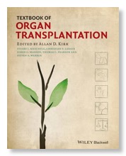
Brought to you by the world’s leading transplant clinicians, Textbook of Organ Transplantation provides a complete and comprehensive overview of modern transplantation in all its complexity, from basic science to gold-standard surgical techniques to post-operative care, and from likely outcomes to considerations for transplant program administration, bioethics and health policy.
Beautifully produced in full color throughout, and with over 600 high-quality illustrations, it successfully
- Provides a solid overview of what transplant clinicians/surgeons do, and with topics presented in an order that a clinician will encounter them.
- Presents a holistic look at transplantation, foregrounding the interrelationships between transplant team members and non-surgical clinicians in the subspecialties relevant to pre- and post-operative patient care, such as gastroenterology, nephrology, and cardiology.
- Offers a focused look at pediatric transplantation, and identifies the ways in which it significantly differs from transplantation in adults.
- Includes coverage of essential non-clinical topics such as transplant program management and administration; research design and data collection; transplant policy and bioethical issues.

Editors are Allan D. Kirk, Stuart J. Knechtle, Christian P. Larsen, Joren C. Madsen, Thomas C. Pearson, and Steven A. Webber.
I.M. Sauer, N. Raschzok und P. Neuhaus contributed chapter 47: „Artificial Liver, In Vivo Tissue Engineering, and Organ Printing – Solutions for Organ Scarcity“

The Wiley-Blackwell book (ISBN: 978-1-118-87014-3, 1880 pages) is available here.
Read More
Decellularization of porcine livers
Stacks Image 34142
Ben Strücker’s paper entitled „Porcine liver decellularization under oscillating pressure conditions – A technical refinement to improve the homogeneity of the decellularization process“ has been accepted for publication in Tissue Engineering, Part C: Methods.
Co-authors are K. Hillebrandt, R. Voitl, A. Butter, R.B. Schmuck, A. Reutzel-Selke, D. Geisel, K. Joehrens, P.A. Pickerodt, N. Raschzok, G. Puhl, P. Neuhaus, J. Pratschke, and I.M. Sauer.

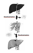
Decellularization and recellularization of parenchymal organs may facilitate the generation of autologous functional liver organoids by repopulation of decellularized porcine liver matrices with induced liver cells. We present an accelerated (7 h overall perfusion time) and effective protocol for human scale liver decellularization by pressure-controlled perfusion with 1% Triton X-100 and 1% SDS via the hepatic artery (120 mmHg) and portal vein (60 mmHg). In addition, we analyzed the effect of oscillating pressure conditions on pig liver decellularization (n=12). The proprietary perfusion device used to generate these pressure conditions mimics intra-abdominal conditions during respiration to optimize microperfusion within livers and thus optimize the homogeneity of the decellularization process. The efficiency of perfusion decellularization was analyzed by macroscopic observation, histological staining (H&E, Sirius red, Alcian blue), immunohistochemical staining (collagen IV, laminin, fibronectin) and biochemical assessment (DNA, collagen, glycosaminoglycans) of decellularized liver matrices. The integrity of the extracellular matrix post-decellularization was visualized by corrosion casting and three-dimensional CT scanning. We found that livers perfused under oscillating pressure conditions (P+) showed a more homogenous course of decellularization and contained less DNA compared to livers perfused without oscillating pressure conditions (P-). Microscopically, livers from the (P-) group showed remnant cell clusters, while no cells were found in livers from the (P+) group. The grade of disruption of the ECM was higher in livers from the (P-) group, although the perfusion rates and pressure did not significantly differ. Immunohistochemical staining revealed that important matrix components were still present after decellularization. Corrosion casting showed an intact vascular (portal vein and hepatic artery) and biliary framework. In summary, the presented protocol for pig liver decellularization is quick (7 h) and effective. The application of oscillating pressure conditions improves the homogeneity of perfusion and thus the outcome of the decellularization process.
Read More
Decellularization and oscillating pressure conditions
Stacks Image 34196
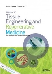
The Journal of Tissue Engineering and Regenerative Medicine accepted our latest paper on „Improved rat liver decellularization by arterial perfusion under oscillating pressure conditions“ for publication. Authors are B. Struecker, A. Butter, K. Hillebrandt, D. Polenz , A. Reutzel-Selke, P. Tang, S. Lippert, A. Leder, S. Rohn, D. Geisel, T. Denecke, K. Aliyev, K. Jöhrens, N. Raschzok, P. Neuhaus, J. Pratschke and I.M. Sauer.

One approach of regenerative medicine to generate functional hepatic tissue in vitro is de- and recellularization and several protocols for the decellularization of different species have been published. To the best of our knowledge this is the first report on rat liver decellularization by perfusion via the hepatic artery under oscillating pressure conditions, intending to optimize microperfusion and minimize damage to the ECM. Four decellularization protocols were compared: perfusion via the portal vein (PV) or the hepatic artery (HA), with (+P) or without (-P) oscillating pressure conditions. All rat livers (n=24) were perfused with 1% Triton X-100 and 1% SDS, each for 90 minutes with a perfusion rate of 5ml/min. Perfusion decellularization was observed macroscopically and the decellularized liver matrices (DLMs) were analyzed by histology and biochemical analyses (e.g., levels of DNA, glycosaminoglycans (GAG), and hepatocyte growth factor (HGF). Livers decellularized via the hepatic artery and under oscillating pressure showed a more homogenous decellularization and less remaining DNA, compared to livers of the other experimental groups. The novel decellularization method described is effective, quick (3 hours) and gentle to the ECM and thus represents an improvement of existing methodology.
Read More
Nature Reviews Gastroenterology and Hepatology
Stacks Image 34211
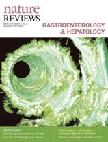
Nature Reviews Gastroenterology and Hepatology invited us to provide a review on liver support strategies.

The treatment of end-stage liver disease and acute liver failure remains a clinically relevant issue. Although orthotopic liver transplantation is a well-established procedure, whole-organ transplantation is invasive and increasingly limited by the unavailability of suitable donor organs. Artificial and bioartificial liver support systems have been developed to provide an alternative to whole organ transplantation, but despite three decades of scientific efforts, the results are still not convincing with respect to clinical outcome. In this Review, conceptual limitations of clinically available liver support therapy systems are discussed. Furthermore, alternative concepts, such as hepatocyte transplantation, and cutting-edge developments in the field of liver support strategies, including the repopulation of decellularized organs and the biofabrication of entirely new organs by printing techniques or induced organogenesis are analysed with respect to clinical relevance. Whereas hepatocyte transplantation shows promising clinical results, at least for the temporary treatment of inborn metabolic diseases, so far data regarding implantation of engineered hepatic tissue have only emerged from preclinical experiments. However, the evolving techniques presented here raise hope for bioengineered liver support therapies in the future.


Update: The review „Liver support strategies: cutting-edge technologies“ (authors: Benjamin Struecker, Nathanael Raschzok & Igor M. Sauer) is now available.
Read More
41st Annual Congress of the ESAO in Rome
Stacks Image 34245
Prof. Celestino Pio Lombardi, Gemelli Polyclinics, Catholic University of the Sacred Heart in Rome, and Prof. Gerardo Catapano organize the 41st Annual Congress of the European Society for Artificial Organs (ESAO) in Rome, Italy.

The 41st ESAO Congress will be focused on “Patient happiness: the Holy Grail of organ substitution”, and will be held on September 17-20, 2014 at the Giovanni XXIII congress center located inside the Gemelli Polyclinics in Rome, Italy. The meeting is expected to attract aprox. 600 participants with both clinical and technical expertise from all over the world.
The objective of the Congress is to bring together scientists with different backgrounds working at the development, optimization and translation to the clinics of treatments of organ deficits based on the use of artificial, bioartificial, and cell-based organs or prostheses, and to discuss the importance of technical, psychological and ethical issues to the happiness of patients with serious tissue or organ deficits so as to re-define the design criteria of devices and treatments.
Read More
12th Congress of the Cell Transplant Society
Stacks Image 34305
The 12th Congress of the Cell Transplant Society tookplace in Milan, Italy, from July 7 to 11, 2013.
Nathanael Raschzok gave a presentation on "Loco-regional detection and stimulation of transplanted liver cells by particle-based miRNA depletion" and Martina Mogl on "isolation of adult hepatocytes and progenitor cells from explanted diseased human livers“.
Read More
IEEE Engineering in Medicine and Biology Society
Stacks Image 34315
The 35th Annual International Conference of the IEEE Engineering in Medicine and Biology Society (EMBC’13) will take place 3-7 July 2013, at the Osaka International Convention Center, in Osaka, Japan.
The conference will cover diverse topics such as biomedical engineering, healthcare technologies, and medical and clinical applications.
The ESAO will be represented via a minisymposium entitled "Artificial Organs for Metabolic Support. The most Challenging Problems". Jan Wojcicki will give a presentation on "Artificial Organs for Metabolic Support: The Most Challenging Problems of Artificial Pancreas", Bernd Stegmayr on "Artificial Organs for Metabolic Support: The Most Challenging Problems in Severe Kidney Injury When Dialysis Is Necessary" and Igor Sauer on "Artificial Organs for Metabolic Support: The Most Challenging Problems of Liver Support".
Read More
Noninvasive monitoring of liver cell transplantation
Stacks Image 34325
Our latest review on „Noninvasive monitoring of liver cell transplantation“ (authors are N. Raschzok, H.M. Morgül, L. Stelter & I.M. Sauer) is available in Imaging in Medicine, 2013; 5: 47-61:
Liver cell transplantation was developed as a therapeutic alternative to solid liver transplantation in the management of liver-based metabolic disorders and may be useful for the treatment of acute or chronic liver failure. While clinical studies have demonstrated temporal amelioration of the symptoms of metabolic liver disorders by transplanted liver cells, the long-term outcome of liver cell transplantation is still insufficient. A major limitation for improving liver cell transplantation is the inability to track the fate of cells once they have been infused. Radionuclide-based imaging, MRI and optical methods have been investigated as methods for noninvasive monitoring of liver cell transplantation. This article summarizes and critically discusses these approaches, with a special focus on MRI-based tracking of transplanted liver cells and provides an outlook on possible clinical applications for the near future.
Read More
Einladunfgzur öffentlichen Abschlusspräsentation
Stacks Image 34335
Sehr geehrte Damen und Herren,
Dear ladies and gentlemen,

Sie sind herzlich eingeladen, unserer öffentlichen Abschlusspräsentation unseres EU/EFRE-Projekts am 20.03.2013 um 15:00 Uhr beizuwohnen. Zusammen mit unseren Partnern der Firma microparticles GmbH werden wir den aktuellen Stand zur „Entwicklung von Partikel zur Detektion und ultralokoregionären Stimulation transplantierter Leberzellen“ darlegen.
You are cordially invited to attend our public presentation of our EU/EFRE project. Together with our partners of microparticles GmbH we will present our latest results concerning the „Development of particles for detection and ultralocoregional stimulation of transplanted liver cells“.

Wann/When?
20.03.2013 um/at 15:00

Wo/Where?
Experimentelle Chirurgie und Regenerative Medizin
Klinik für Allgemein-, Viszeral- und Transplantationschirurgie
Charité, Campus Virchow-Klinikum
Forschungshaus/BMFZ
Pilzraum, 1.OG

Wir wären dankbar, wenn Sie Ihr Kommen via email (anja.selke@charite.de)bestätigen könnten.
RSVP via email (anja.selke@charite.de).
Read More
David Mücke successfully defended thesis
Today, David Mücke successfully defended his thesis magna cum laude with respect to his work on the in vitro evaluation of MRI contrast agents for detection of primary human hepatocytes („In vitro Evaluierung von Magnetresonanztomografie-Kontrastmitteln für die Markierung primärer humaner Hepatozyten“).

Congratulations !
Read More
XXXX ESAO Congress in Glasgow
Stacks Image 34383
The European Society for Artificial Organs (ESAO) invites you to the XXXX ESAO Congress of the society to be held in Glasgow (Scotland, UK), September 11th-14th 2013.The motto of the ESAO congress will be 'lab to patient - from concept to treatment.
Read More
Presentations at EASO 2012 in Rostock, Germany
Stacks Image 34390
This year members of the team gave the following presentations:

“Liver: Current regenerative strategies and future solutions for the liver" (N. Raschzok, oral presentation)
"Neohybrid liver graft - a novel concept of in vivo tissue-engineering" (S. Rohn, oral presentation)
"Micro RNAs in liver regeneration: the mysterious MIR-352" (L. Lisboa, oral presentation)
"Micron-sized iron oxide particles for detection and loco-regional stimulation of transplanted liver cells" (A. Leder, oral presentation)
"Prospective validation of cross organ serum protein biomarkers – Initial results with CXCL9 and CD44 for diagnosis of acute liver rejection" (K.A. Prabowo, poster presentation)
Read More
MRI and ectopic liver cell transplantation - new paper
Stacks Image 34403
Nathanael Raschzok’s latest paper on „Feasibility of fast dynamic MRI for noninvasive monitoring during ectopic liver cell transplantation to the spleen in a porcine model“ is now available in AJR Am J Roentgenol. 2012 Jun;198(6):1417-23.
Liver cell transplantation is a promising approach for the treatment of metabolic liver disorders. However, a method for noninvasive monitoring during liver cell transplantation is not available clinically. The aim of this study was to investigate the feasibility of fast dynamic MRI monitoring during liver cell infusion to the spleen, which is considered an ectopic implantation site for liver cell transplantation. Porcine liver cells were labeled with micron-sized iron oxide particles and infused to the spleens of pigs (n = 5) via the lineal artery. MRI was performed using a 3-T MR scanner. Initially, T1- and T2-weighted pulse sequences were tested. Thereafter, fast dynamic MRI was performed during cell infusion. MR findings were verified by immunohistological examinations.

Images from static MRI (TR/TE, 2500/105.2) showed significantly lower signal intensity and signal-to-noise ratio after cell infusion compared with pretransplant images. T2-weighted fast dynamic MRI enabled visualization of signal decrease of the spleen during cell infusion. When cells were infused systemically, no signal changes in the spleen were observed. This study shows that fast dynamic MRI can enable noninvasive monitoring during liver cell transplantation to the spleen. This approach could be useful for preclinical studies and for quality control of clinical liver cell transplantation.
Read More
TTS 2012 - Thank you!
Stacks Image 34417

The Science Circuses are located at each end of the Exhibition Area. You will find the Mini-Theatres for the Mini-Oral Sessions here as well as the Poster Lounges and the Web Stations. Science Circus I includes Mini-Theatres 1 – 5; Science Circus II includes Mini-Theatres 6 – 10. In each Science Circus you will find Web Stations where you can access the web as well as the ePosters.
Via wireless headphones and dedicated channels for each mini-theater this is – to our knowledge – the first successful concept for mini-oral presentations!
Thanks to m-events and Interplan for the tremendous support!

Read More
TTS 2012 | Over 500 presentations online!

The LOC would like to thank all participants for a tremendous 24th International Congress of The Transplantation Society in Berlin!
2268 abstracts were submitted and more than 4800 delegates from 94 countries attended the congress!
The Postgraduate Weekend offered 14 workshops with 45 speakers. Beside 29 Sunrise Symposia with 103 Speakers, 34 State of the Art Sessions with 138 speakers and five Plenary Sessions (16 speakers) we had 53 sessions with 465 oral presentations and 37 sessions with 287 mini oral presentations !
Furthermore, we would like to thank Astrid Enke, Lena Dochat and Stefanie Rensch (Interplan) and the technical experts at m-Events for their excellent work !
As Science and Medicine is nothing without vivid life we would like to thank Rotfront, Berlin Comedian Harmonists and the Capital Dance Orchestra for their memorable performances!

All the great photos by Jan Pauls.
The organ sculptures were made by Jan Pareike.

Read More
<<  Page 6 / 8  >>
Year
© 2025 Prof. Dr. Igor M. Sauer | Charité - Universitätsmedizin Berlin | Disclaimer

This website or its third-party tools use cookies, which are necessary to its functioning and required to achieve the purpose illustrated in the Disclaimer. By closing this banner, scrolling this page, clicking a link or continuing to browse otherwise, you agree to the use of cookies.