News

Decellularization of porcine livers
Stacks Image 25053
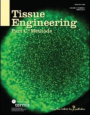
Ben Strücker’s paper entitled „Porcine liver decellularization under oscillating pressure conditions – A technical refinement to improve the homogeneity of the decellularization process“ has been accepted for publication in Tissue Engineering, Part C: Methods.
Co-authors are K. Hillebrandt, R. Voitl, A. Butter, R.B. Schmuck, A. Reutzel-Selke, D. Geisel, K. Joehrens, P.A. Pickerodt, N. Raschzok, G. Puhl, P. Neuhaus, J. Pratschke, and I.M. Sauer.

Decellularization and recellularization of parenchymal organs may facilitate the generation of autologous functional liver organoids by repopulation of decellularized porcine liver matrices with induced liver cells. We present an accelerated (7 h overall perfusion time) and effective protocol for human scale liver decellularization by pressure-controlled perfusion with 1% Triton X-100 and 1% SDS via the hepatic artery (120 mmHg) and portal vein (60 mmHg). In addition, we analyzed the effect of oscillating pressure conditions on pig liver decellularization (n=12). The proprietary perfusion device used to generate these pressure conditions mimics intra-abdominal conditions during respiration to optimize microperfusion within livers and thus optimize the homogeneity of the decellularization process. The efficiency of perfusion decellularization was analyzed by macroscopic observation, histological staining (H&E, Sirius red, Alcian blue), immunohistochemical staining (collagen IV, laminin, fibronectin) and biochemical assessment (DNA, collagen, glycosaminoglycans) of decellularized liver matrices. The integrity of the extracellular matrix post-decellularization was visualized by corrosion casting and three-dimensional CT scanning. We found that livers perfused under oscillating pressure conditions (P+) showed a more homogenous course of decellularization and contained less DNA compared to livers perfused without oscillating pressure conditions (P-). Microscopically, livers from the (P-) group showed remnant cell clusters, while no cells were found in livers from the (P+) group. The grade of disruption of the ECM was higher in livers from the (P-) group, although the perfusion rates and pressure did not significantly differ. Immunohistochemical staining revealed that important matrix components were still present after decellularization. Corrosion casting showed an intact vascular (portal vein and hepatic artery) and biliary framework. In summary, the presented protocol for pig liver decellularization is quick (7 h) and effective. The application of oscillating pressure conditions improves the homogeneity of perfusion and thus the outcome of the decellularization process.
Back
Stacks Image 27655
Our manuscript "Depletion of donor dendritic cells ameliorates immunogenicity of both skin and hind limb transplants" has been accepted for publication in Frontiers in Immunology, section Alloimmunity and Transplantation. Authors are Muhammad Imtiaz Ashraf, Joerg Mengwasser, Anja Reutzel-Selke, Dietrich Polenz, Kirsten Führer, Steffen Lippert, Peter Tang, Edward Michaelis, Rusan Catar, Johann Pratschke, Christian Witzel, Igor M. Sauer, Stefan G. Tullius, and Barbara Kern.

Acute cellular rejection remains a significant obstacle affecting successful outcomes of organ transplantation including vascularized composite tissue allografts (VCA). Donor antigen presenting cells (APC), particularly dendritic cells (DC), orchestrate early alloimmune responses by activating recipient effector T cells. Employing a targeted approach, we investigated the impact of donor-derived conventional DC (cDC) and APC on the immunogenicity of skin and skin-containing VCA grafts, using mouse models of skin and hind limb transplantation.
By post-transplantation day 6, skin grafts demonstrated severe rejections, characterized by predominance of recipient CD4 T cells. In contrast, hind limb grafts showed moderate rejection, primarily infiltrated by CD8 T cells. While donor depletion of cDC and APC reduced frequencies, maturation, and activation of DC in all analysed tissues of skin transplant recipients, reduction in DC activities was only observed in the spleen of hind limb recipients. Donor cDC and APC depletion did not impact all lymphocyte compartments but significantly affected CD8 T cells and activated CD4 T in lymph nodes of skin recipients. Moreover, both donor APC and cDC depletion attenuated the Th17 immune response, evident by significantly reduced Th17 (CD4+IL-17+) cells in the spleen of skin recipients and reduced levels of IL-17E and lymphotoxin-α in the serum samples of both skin and hind limb recipients. In conclusion, our findings underscore the highly immunogenic nature of skin component in VCA. The depletion of donor APC and cDC mitigates the immunogenicity of skin grafts while exerting minimal impact on VCA.

Archive


Categories

Year

This website or its third-party tools use cookies, which are necessary to its functioning and required to achieve the purpose illustrated in the Disclaimer. By closing this banner, scrolling this page, clicking a link or continuing to browse otherwise, you agree to the use of cookies.