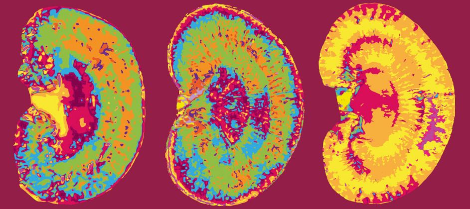
Wibke Schulte: BIH Charité Clinician Scientist
Stacks Image 24516
Dr. med. Wibke Schulte successfully applied for the BIH Charité Clinician Scientist Program with her project „Die Rolle von MIF in der humanen akuten Peritonitis | Role of MIF in human acute peritonitis“. Mentors are Priv.-Doz. Dr. med. Felix Aigner and Prof. Dr. Igor M. Sauer.
Read More
Isolation of primary human hepatocytes & LiMax-test
Stacks Image 24537
Tissue Engineering (Part C: Methods) accepted our paper entitled "The predictive value of the LiMAx-test for the isolation of primary human hepatocytes".
Authors are R.D. Major, M. Kluge, M. Jara, M. Nösser, R. Horner, J. Gassner, B. Struecker, P. Tang, S. Lippert, A. Reutzel-Selke, D. Geisel, T. Denecke, M. Stockmann, J. Pratschke, I.M. Sauer, and N. Raschzok.

The need for primary human hepatocytes is constantly growing, for basic research as well as for therapeutic applications. However, the isolation outcome strongly depends on the quality of liver tissue, and we are still lacking a preoperative test that allows the prediction of the hepatocyte isolation outcome. Here we evaluated the “maximal liver function capacity test” (LiMAx) as predictive test for the quantitative and qualitative outcome of hepatocyte isolation. This test is already used in clinical routine to measure preoperative and to predict postoperative liver function.
The patient’s preoperative mean LiMAx was obtained from the patient records and preoperative CT and MRI images were used to calculate the whole liver volume in order to adjust the mean LiMAx. The outcome parameters of the hepatocyte isolation procedures were analyzed in correlation with the adjusted mean LiMAx.
Primary human hepatocytes were isolated from partial hepatectomies (n=64).
From these 64 hepatectomies we included 48 to our study and correlated their isolation outcome parameters with volume corrected LiMAx values. From a total of 11 hepatocyte isolation procedures, metabolic parameters (albumin, urea and aspartate aminotransferase) were assessed during the hepatocyte cultivation period of 5 days. The volume adjusted mean LiMAx showed a significant positive correlation with the total cell yield (p= 0.049;r= 0.242;n= 48). The correlations of volume adjusted LiMAx values with viable cell yield and cell viability did not reach statistical significance. A sub-group analysis of isolations from patients with colorectal metastasis revealed a significant correlation between volume adjusted mean LiMAx and total cell yield (p= 0.012;r= 0.488;n= 21) and viable cell yield (p=0.034;r=0.405;n=21). Whereas a sub-group analysis of isolations of patients with carcinoma of the biliary tree showed significant correlations of volume adjusted mean LiMAx with cell viability (r= 0.387;p= 0.046;n=20) and lacked significant correlations with total cell yield (r= - 0.060;p= 0.401;n=20) and viable cell yield (r= 0.012;p= 0.480;n=20). The volume-adjusted mean LiMAx did not show a significant correlation with any of the metabolic parameters. In conclusion, the LiMAx-test might be a useful tool to predict the quantitative outcome of hepatocyte isolation, as long as underlying liver disease is taken into consideration.
Read More
Barbara Kern: BIH Charité Clinician Scientist
Stacks Image 24548
Dr. Barbara Kern successfully applied for the BIH Charité Clinician Scientist Program. With her project "Novel Treatment and Diagnostic Approaches Utilizing the Role of Dendritic Cells in Immune Responsivness" Barbara will be engaged in our Vascular Tissue Allotransplantation Initiative (Project VCA). The Clinician Scientist Program is a modern career pathway within academic medicine that allows physicians to pursue a structured residency with time set aside for clinical and basic research. At the end of the program, participants will have completed their residency and, ideally, their postdoctoral teaching qualification (Habilitation). The program is intended to produce a new generation of scientists with translational training who will help speed up the rate at which scientific findings are translated into application.
Read More
Future Medicine 2017
Stacks Image 24555
What’s trending? What’s new in health science? To find out, please save the date for the second Future Medicine in Berlin on November 7, 2017. Tagesspiegel and Berlin Institute of Health, together with Charité - Universitätsmedizin Berlin and Max Delbrück Center for Molecular Medicine, will feature outstanding international scientists, great visions of the future of medicine, and an exceptional concentration of knowledge. The four sessions of Future Medicine 2017 will be: 

Digital Health and Big Data
Precision Medicine and Predictive Models
Cell and Gene Therapies
Stem Cells and Human Disease Modeling  

Simon Moosburner, a student of medicine interested in regenerative medicine and future technologies, will talk about “Virtual & Mixed Reality: The Next Milestone in Surgery?” at Future Medicine 2017.
Read More
21. Chirurgischen Forschungstage
Stacks Image 24570
The 21. Chirurgische Forschungstage took place in Cologne. Five of our students gave terrific presentations: S. Moosburner gave an oral presentation on „Steatotic Liver Transplantation – a Growing Problem with Severe Complications“, H. Everwien on „Different biological scaffolds as a platform for engineering an endocrine Neo-Pancreas by using decellularization and recellularization techniques“, M. Noesser on „A comprehensive description of the development of a stable closed circuit for ex vivo rat liver machine perfusion“, R. Horner on „Is Percoll purifcation necessary for isolation of primary human hepatocytes?“, and N. Seiffert on „Recellularization of Decellularized Bovine Carotid Arteries using Human Endothelial Progenitor Cells: One Step towards an Autologous Bypass Graft“.
Read More
ESOT The Future of Transplantation
Stacks Image 24587
The ESOT YPT Workshop and its Presidential Debate gives you a chance to engage with a variety of speakers in a lively ‘town hall’ format, as well as connecting with other young transplant professionals from around the world. The session will begin with talks from three key presenters on the past, present, and future of transplantation and open Q&As with each presenter. 

09:00– 10:35 - The future of transplantation: 3 lectures on Past, Present and Future CHAIRS: Alice Koenig, Lyon, France   Thomas Resch, Innsbruck, Austria
09:05 Lecture: The future of transplantation - Historical Perspective Sir Roy Calne, Cambridge, United Kingdom 09:25 Open Q&A with Roy Calne
09:35 Lecture: The future of transplantation - Present perspective Jan Lerut, Brussels, Belgium 09:55 Open Q&A with Jan Lerut
10:05 Lecture: The future of transplantation - Future perspective  Igor Sauer, Berlin, Germany
10:25 Open Q&A with Igor Sauer
Read More
Mixed Reality in Visceral Surgery
Stacks Image 24597
Annals of Surgery accepted our manuscript "Mixed Reality in visceral surgery - Development of a suitable workflow and evaluation of intraoperative use-cases" for publication. The paper evaluates the application of a mixed reality (MR) head-mounted display (HMD) for the visualization of anatomical structures in complex visceral-surgical interventions. A workflow was developed and technical feasibility was evaluated. 
Medical images are still not seamlessly integrated into surgical interventions and thus, remain separated from the surgical procedure. Surgeons need to cognitively relate two-dimensional sectional images to the three-dimensional (3D) during the actual intervention. MR applications simulate 3D images and reduce the offset between working space and visualization allowing for improved spatial-visual approximation of patient and image. The surgeon’s field of vision was superimposed with a 3D-model of the patient’s relevant liver structures displayed on a MR-HMD. This set-up was evaluated during open hepatic surgery. A suitable workflow for segmenting image masks and texture mapping of tumors, hepatic artery, portal vein and the hepatic veins was developed. The 3D model was positioned above the surgical site. Anatomical reassurance was possible simply by looking up. Positioning in the room was stable without drift and minimal jittering. Users reported satisfactory comfort wearing the device without significant impairment of movement. MR technology has high potential to improve the surgeon’s action and perception in open visceral surgery by displaying 3D anatomical models close to the surgical site. Superimposing anatomical structures directly onto the organs within the surgical site remains challenging since the abdominal organs undergo major deformations due to manipulation, respiratory motion and the interaction with the surgical instruments during the intervention. A further application scenario would be intraoperative ultrasound examination displaying the image directly next to the transducer. Displays and sensor-technologies as well as biomechanical modeling and object-recognition algorithms will facilitate the application of MR-HMD in surgery in the near future. Authors are I.M. Sauer, M. Queisner, P. Tang, S. Moosburner, O. Hoepfner, R. Horner, R. Lohmann and J. Pratschke.
Read More
Cells isolated from diseased explanted livers
Stacks Image 24617
The International Journal of Artificial Organs (official journal of the European Society for Artificial Organs [ESAO]) published our paper on Isolation, characterization and cold storage of cells isolated from diseased explanted livers. Authors are Belaschk E, Rohn S, Mukiibi R, Reutzel-Selke A, Tang P, Sawitzki B, Pratschke J, Sauer IM and Mogl MT.

Livers discarded after standard organ retrieval are commonly used as a cell source for hepatocyte transplantation. Due to the scarcity of organ donors, this leads to a shortage of suitable cells for transplantation. Here, the isolation of liver cells from diseased livers removed during liver transplantation is studied and compared to the isolation of cells from liver specimens obtained during partial liver resection. Hepatocytes from 20 diseased explanted livers (Ex-group) were isolated, cultured and stored at 4°C for up to 48 hours, and compared to hepatocytes isolated from the normal liver tissue of 14 liver lobe resections (Rx-group). The nonparenchymal cell fraction (NPC) was analyzed by flow cytometry to identify potential liver progenitor cells, and OptiPrep™ (Sigma-Aldrich) density gradient centrifugation was used to enrich the progenitor cells for immediate transplantation. There were no differences in viability, cell integrity and metabolic activity in cell culture and survival after cold storage when comparing the hepatocytes from the Rx-group and the Ex-group. In some cases, the latter group showed tendencies of increased resistance to isolation and storage procedures. The NPC of the Ex-group livers contained considerably more EpCAM+ and significantly more CD90+ cells than the Rx-group. Progenitor cell enrichment was not sufficient for clinical application. Hepatocytes isolated from diseased explanted livers showed the essential characteristics of being adequate for cell transplantation. Increased numbers of liver progenitor cells can be isolated from diseased explanted livers. These results support the feasibility of using diseased explanted livers as a cell source for liver cell transplantation.
Read More
ECRT - Advanced Scientist Grant
Stacks Image 24627
PD Dr. Nathanael Raschzok receives one of the 2017 Einstein Center for Regenerative Therapies (ECRT) Kickbox – Advanced Scientist Grant.
Einstein Center for Regenerative Therapies Kickbox – advanced scientist grant. The project is entitled „Overcoming steatotic compromise – Reconstitution of endogenous repair in severely steatotic liver grafts by metabolic reconditioning“. The project will be conducted by Nathanael Raschzok, Angelika Kusch, Duska Dragun, and Igor M. Sauer.

In order to stimulate excellent and creative research ideas that might take regenerative therapies a vital step forward, the Einstein Center offers a special two-stage funding scheme. At first, the Kickbox seed grant provides a great framework to investigate initial ideas and to develop sound research concepts. Subsequently, the flexible funds enable the realisation of projects that evolved from the Kickbox initiation phase in order to reach the scientific goals of the Einstein Center. Congratulations!
Read More
Magnetic field and cells labeled with IO particles
Stacks Image 24637
Our paper entitled "The magnetic field of magnetic resonance imaging systems does not affect cells labeled with micrometer-sized iron oxide particles," has been accepted for publication in Tissue Engineering, Part C: Methods. Authors are Martin Kluge, Annekatrin Leder, Karl H. Hillebrandt, Benjamin Struecker, Dominik Geisel, Timm Denecke, Rebeka D. Major, Anja Reutzel-Selke, Peter Tang, Steffen Lippert, Christian Schmidt, Johann Pratschke, Igor M. Sauer, and Nathanael Raschzok.

Labeling using iron oxide particles enables cell tracking via magnetic resonance imaging (MRI). However, the magnetic field can affect the particle-labeled cells. Here, we investigated the effects of a clinical MRI system on primary human hepatocytes labeled using micrometer-sized iron oxide particles (MPIOs).  HuH7 tumor cells were incubated with increasing concentrations of biocompatible, silica-based, micron-sized iron oxide-containing particles (sMPIO; 40 – 160 particles/cell). Primary human hepatocytes were incubated with 100 sMPIOs/cell. The particle-labeled cells and the native cells were imaged using a clinical 3.0-T MRI system, whereas the control groups of the labeled and unlabeled cells were kept at room temperature without exposure to a magnetic field. Viability, formation of reactive oxygen species, aspartate aminotransferase leakage, and urea and albumin synthesis were assessed over a culture period of 5 days. 
The dose finding study showed no adverse effects of the sMPIO labeling on HuH7 cells. MRI had no adverse effects on the morphology of the sMPIO-labeled primary human hepatocytes. Imaging using the T1- and T2-weighted sequences did not affect the viability, transaminase leakage, formation of reactive oxygen species, or metabolic activity of the sMPIO-labeled cells or the unlabeled, primary human hepatocytes. sMPIOs did not induce adverse effects on the labeled cells under the conditions of the magnetic field of a clinical MRI system.
Read More
Charité BIH Entrepreneurship Summit
Stacks Image 24647

The Charité BIH Entrepreneurship Summit is a preeminent international cross-disciplinary forum for sharing and exploring the most important discoveries and emerging trends influencing the future of healthcare around the world.

Every year in May over 400 global leaders in healthcare innovation, including entrepreneurs, scientists, physicians, investors, policymakers, and business leaders convene at the Charité BIH Entrepreneurship Summit in the vibrant German capital of Berlin. We offer our participants a one-of-a-kind opportunity to meet with world-class specialists working at Charité & MDC, to build relationships with prominent international partners and experts working in the healthcare industry, and to help grow businesses.

This year's 10th Charité Entrepreneurship Summit will again take place at the Berlin-Brandenburg Academy of Sciences and Humanities on May 8 - 9, 2017. The Summit is significantly supported by the Berlin Institute of Health and focusses on 'Global Challenges of Healthcare'. Israel will be the official partner country for the Summit 2017. We are looking forward to learning more about the Israeli innovation & start-up culture, funding opportunities and challenges in Healthcare.

The Summit's two-day agenda will address a wide variety of topical issues including change of innovation culture, healthy aging & degenerative diseases, virtual reality and mental health. In addition, the Summit will seek to engage participants in lively discussions about business, science and the intersection of the two. Startups and Entrepreneurs are invited to apply for the LifeSciences VentureMarket, a platform to present their companies to a pool of international angels, venture investors, and corporate funds at this year's Summit.

Read More
BIH Paper of the Month
Stacks Image 24659
Benjamin Strücker, Hendrik Napierala and the rest of the team were awarded with the BIH Paper of the Month for their publication on a new method for developing a transplantable endocrine Neo-Pancreas.

The BIH Paper of the Month is awarded by the BIH Board of Directors to honor a special publication achievement from the joint research space of Charité and MDC. The Paper of the Month is sponsored by the Stiftung Charité as part of its Johanna Quandt Private Excellence Initiative. 

H. Napierala, K.-H. Hillebrandt, N. Haep, P. Tang, M. Tintemann, J. Gassner, M. Noesser, H. Everwien, N. Seiffert, M. Kluge, E. Teegen, D. Polenz, S. Lippert, D. Geisel, A. Reutzel Selke, N. Raschzok, A. Andreou, J. Pratschke, I. M. Sauer & B. Struecker. Engineering an endocrine Neo-Pancreas by repopulation of a decellularized rat pancreas with islets of Langerhans. Scientific Reports 7. Article number: 41777 (2017) doi:10.1038/srep41777
Read More
ECRT Kickbox - Junior Scientist Grant
Stacks Image 24674
Karl Hillebrandt receives one of the 2017 Einstein Center for Regenerative Therapies (ECRT) Kickbox – Junior Scientist Grant. The project is entitled "Fighting liver cirrhosis? Establishment and analysis of decellularized human cirrhotic liver slices as a 3-dimensional model to study cell matrix interactions".

Liver cirrhosis is one of the main indications for liver transplantation. Due to the organ shortage, this therapy option is limited to the minority of patients suffering from cirrhosis. Therefore, there is a need of alternative treatment options.The aim of our project is to establish a decellularization protocol for human cirrhotic livers slices, which preserves the natural extracellular matrix (ECM) of cirrhotic livers. These decellularized liver slices will serve as a 3 dimensional model to study cell matrix interactions. If we are able to establish a protocol which will preserve the ECM, we will conduct in vitro recellularization experiments to study how the cirrhotic ECM will change the genotype and phenotype of different cell types. With this knowledge we aim to modify specific cell types in vivo or vitro for example prior to cell transplantation. Our ambition is to steer the cell matrix interaction via these modified cells after their transplantation and thereby halt or even reverse the progress of liver cirrhosis. This approach may offer an alternative treatment option in the future.

Team : Karl Hillebrandt, Oliver Klein, Ben Strücker, Igor Sauer  
Read More
Einstein Visiting Fellow Prof. Stefan G. Tullius
Stacks Image 24689

Prof. Dr. Stefan G. Tullius, Harvard Medical School, became Einstein BIH Visiting Fellow at the Charité, Department of Surgery. 

Vascular Composite Tissue Allotransplantation (VCA) has developed from a pioneering endevour to a clinical reality during the last 15 years. More than 150 extremity, face, knee, and most recently uterus and penis transplants have been performed during the last 15 years. VCA activities have been seen around the globe. Although feasibility of the procedure has been shown, it is still emerging and far from being a standard clinical procedure. 

The Charité has been an international leader in transplantation research for decades. However, neither VCA basic clinical research programs are currently offered in Berlin or Germany in a meaningful way.The involvement of Prof. Tullius as an Einstein BIH Visiting Fellow is expected to synergize and accelerate efforts igniting both a clinical research transplant program and a basic research group of excellence. The overall objective of our integrated basic and clinical research working group Vascular Composite Tissue Transplantation has three main goals:

To establish a basic Research group of Excellence: VCAs offer unique opportunities to study novel aspects of antigenicity, immune modulation and neo-vascularization. One important aspect that distinguishes VCA from solid organ transplants is their blood supply through a vascularized arterial in- and venous outflow in addition to sprouting new vessels at the large interface of VCA with recipient tissue.

To establish a clinical bio-repository as a pre-requisite for a clinical research VCA program.

To implement a clinical VCA research program (hand, abdominal wall, uterus) as a multi- disciplinary and translational effort.

Read More
<<  Page 7 / 12  >>
© 2025 Prof. Dr. Igor M. Sauer | Charité - Universitätsmedizin Berlin | Disclaimer

This website or its third-party tools use cookies, which are necessary to its functioning and required to achieve the purpose illustrated in the Disclaimer. By closing this banner, scrolling this page, clicking a link or continuing to browse otherwise, you agree to the use of cookies.