News

Cytokine-armed vaccinia virus promotes cytotoxicity towards pancreatic carcinoma cells
Stacks Image 27891
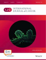
The manuscript "Cytokine-armed vaccinia virus promotes cytotoxicity towards pancreatic carcinoma cells via activation of human intermediary CD56dimCD16dim natural killer cells" by Ruonan Wang, Mengwen Hu, Isis Lozzi, Cao Z.J. Jin, Dou Ma, Katrin Splith, Jörg Mengwasser, Vincent Wolf, Linda Feldbrügge, Peter Tang, Lea Timmermann, Karl H. Hillebrandt, Marieluise Kirchner, Philipp Mertins, Georg Hilfenhaus, Christopher Neumann, Thomas Kammertoens, Johann Pratschke, Thomas Malinka, Igor Sauer, Elfriede Nössner, Zhongsheng Guo and Matthäus Felsenstein has been accepted for publication in the International Journal of Cancer.
 
Pancreatic ductal adenocarcinoma (PDAC) remains a particularly aggressive disease with few effective treatments. The PDAC tumor immune microenvironment (TIME) is known to be immune suppressive. Oncolytic viruses can increase tumor immunogenicity via immunogenic cell death(ICD). We focused on tumor-selective (vvDD) and cytokine-armed Western-Reserve vaccinia viruses (vvDD-IL2, vvDD-IL15) and infected carcinoma cell lines as well as patient-derived primary PDAC cells. In co-culture experiments, we investigated the cytotoxic response and the activation of human natural killer cells (NK). Infection and virus replication were assessed by measuring virus encoded YFP. We then analyzed intracellular signaling processes and oncolysis via in-depth proteomic analysis, immunoblotting and TUNEL assay. Following the co-culture of mock or virus infected carcinoma cell lines with allogenic PBMCs or NK cell lines, CD56+ NK cells were analyzed with respect to their activation, cytotoxicity and effector function. Both, dose- and time-dependent release of danger signals following infection was assayed. Viruses effectively entered PDAC cells and emitted YFP signals. Infection resulted in concomitant oncolysis. The proteome showed reprogramming of normally active core signaling pathways in PDAC occurred(e.g. MAPK-ERK signaling). Danger-associated molecular patterns were released upon infection and stimulated co-cultured NK cells for enhanced effector cytotoxicity. NK cell subtyping revealed enhanced numbers and activation of a rare CD56dimCD16dim population. Tumor cell killing was primarily triggered via Fas ligands rather than granule release, resulting in marked apoptosis. Cytokine-armed vaccinia viruses induced NK cell activation and enhanced cytotoxicity towards human PDAC cells in vitro. The cytokine-armed virus targets the carcinoma cells with great potential to modulate the TIME in PDAC.
Induced pluripotent stem cell (iPSC) line (EXSURGi001-A) from a patient homozygous for the p.Ala165Thr mutation in the MTARC1 gene
Stacks Image 27839
The paper "Induced pluripotent stem cell (iPSC) line (EXSURGi001-A) from a patient homozygous for the p.Ala165Thr mutation in the MTARC1 gene" in Stem Cell Research is available open access. Authors are Peter Tang, Eriselda Keshi, Silvana Wilken, Louise Wutsdorff, Julienne Mougnekabol, Johann Pratschke, Igor M. Sauer and Nils Haep.

Metabolic dysfunction-associated fatty liver disease (MAFLD), the leading cause of end-stage liver disease in developed countries, is expected to increase over the next decade. Characterized by hepatic steatosis, MAFLD is commonly studied in animal models.
Here, we generated a human induced pluripotent stem cell (iPSC) line from a patient homozygous of the protective MTARC1 gene variant rs2642438:A.
This line displays a normal karyotype and typical pluripotent stem cell morphology and can differentiate into all three germ layers in vitro.
Extracellular NAD+ Response to Post-Hepatectomy Liver Failure: Bridging Preclinical and Clinical Findings
Stacks Image 27844
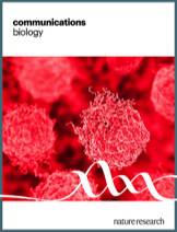
Our manuscript entitled "Extracellular NAD+ Response to Post-Hepatectomy Liver Failure: Bridging Preclinical and Clinical Findings" has been accepted for publication in Communications Biology. Authors are Can Kamali, Philipp Brunnbauer, Kaan Kamali, Al-Hussein Saqr, Alexander Arnold, Gulcin Harman Kamali, Julia Babigian, Eriselda Keshi, Raphael Mohr, Matthäus Felsenstein, Simon Moosburner, Karl Hillebrandt, Jasmin Bartels, Igor Sauer, Frank Tacke, Moritz Schmelzle, Johann Pratschke, and Felix Krenzien.

Liver fibrosis progressing to cirrhosis is a major risk factor for liver cancer, impacting surgical treatment and survival. Our study investigates extracellular Nicotinamide adenine dinucleotide (eNAD+) in liver fibrosis, analyzing patients undergoing surgery and exploring NAD+'s therapeutic potential in a mouse model of extended liver resection and in vitro using 3D hepatocyte spheroids.

eNAD+ correlated with aspartate transaminase (AST) and bilirubin after liver resection (AST: r = 0.2828, p = 0.0087; Bilirubin: r = 0.2584, p = 0.0176). Post-hepatectomy liver failure (PHLF) was associated with higher eNAD+ peaks (n = 10; p = 0.0063). Postoperative eNAD+ levels decreased significantly (p < 0.05), but in advanced liver fibrosis or cirrhosis, this decline diminished or increased. NAD+ biosynthesis enzymes, NAMPT and NMNAT3, were significantly upregulated in higher fibrosis stages (p < 0.0001). NAD+ administration in 3D hepatocyte spheroids rescued hepatocytes from TNFα-induced cell death and improved viability (p < 0.0001). In mice, NAD+ treatment significantly improved survival (p = 0.0155) and liver regeneration (p = 0.0186) after extended liver resection.

eNAD+ is upregulated in PHLF, and NAD+ biosynthesis enzymes show higher expression in liver fibrosis. eNAD+ administration improved survival and hepatocyte viability, offering a potential target for future therapies.

Viscoelastic properties of colorectal liver metastases reflect the tumour cell viability
Stacks Image 27810
Our paper on "Viscoelastic properties of colorectal liver metastases reflect the tumour cell viability" has been accepted for publication in Journal of Translational Medicine.

Colorectal cancer is one of the third most common cancers in the world and up to 50% of the patients develop liver metastases (CRLM) within five years. To improve and personalize therapeutic strategies, new diagnostic tools are urgently needed. An improvement could be achieved by considering biomechanical tumour properties with the implementation of magnetic resonance elastography (MRE). Our main hypothesis is that ex vivo MRE combined with histological evaluation of CRLM could provide the knowledge for using tissue mechanical properties as a diagnostic marker for cell viability in tumours.

We examined 34 CRLM samples from patients who had undergone liver resection at the Charité – Universitätsmedizin Berlin, Department of Surgery. The samples were investigated with an ex vivo MRE.  We employed a frequency range from 500 Hz to 5300 Hz, with increments of 400 Hz. For histological analysis, the samples were stained with H&E for categorization by a board-certified pathologist based on their grade of regression. The radiological response was evaluated using the RECIST-criteria.

Five samples showed major response to chemotherapy, 6 samples partial response, and 23 samples showed no response. Analysis of shear wave speed c significant correlation for frequencies including 2100 Hz and above depending on the grade of regression, indicating that low cell viability in CRLM is associated with higher tumour stiffness. Analysis of frequency-independent values of the SP-model showed a more elastic-solid behaviour at low cell viability. Our results suggest that MRE can be used to characterize the biomechanical properties associated with cell viability in CRLM, showing a higher stiffness and elastic-solid behaviour with high regression. In the future, MRE could help to improve the diagnostic tools to create an individual, tailored therapy plan for patients with CRLM.

Authors are Lisa-Marie Skrip, Simon Moosburner, Peter Tang, Jing Guo, Steffen Görner, Heiko Tzschätzsch, Clarissa Hosse, Uli Fehrenbach, Alexander Arnold, Dominik Modest, Felix Krenzien, Wenzel Schöning, Thomas Malinka, Johann Pratschke, Björn Papke, Josef A. Käs, Ingolf Sack, Igor M. Sauer, and Karl H. Hillebrandt,
Distinctive protein expression in elderly livers in a Sprague-Dawley rat model of normothermic ex vivo liver machine perfusion
Stacks Image 27834
Our manuscript “Distinctive protein expression in elderly livers in a Sprague-Dawley rat model of normothermic ex vivo liver machine perfusion” has been published in the latest issue of the European Journal of Medical Research,
Authors are Maximilian Zimmer, Karl H. Hillebrandt, Nora M. Roschke, Steffen Lippert, Oliver Klein, Grit Nebrich, Joseph M.G.V. Gassner, Felix Strobl, Johann Pratschke, Felix Krenzien, Igor M. Sauer, Nathanael Raschzok, and Simon Moosburner.

Liver grafts are frequently declined due to high donor age or age mismatch with the recipient. To improve the outcome of marginal grafts, we aimed to characterize the performance of elderly vs. young liver grafts in a standardized rat model of normothermic ex vivo liver machine perfusion (NMP).

Livers from Sprague-Dawley rats aged 3 or 12 months were procured and perfused for 6 h using a rat NMP system or collected as a reference group (n = 6/group). Tissue, bile, and perfusate samples were used for biochemical, and proteomic analyses.

All livers cleared lactate during perfusion and continued to produce bile after 6 h of perfusion (614 mg/h). Peak urea levels in 12-month-old animals were higher than in younger animals. Arterial and portal venous pressure, bile production and pH did not differ between groups. Proteomic analysis identified a total of 1477 proteins with oxidoreductase and catalytic activity dominating the gene ontology analysis. Proteins such as aldehyde dehydrogenase 1A1 and 2-Hydroxyacid oxidase 2 were significantly more present in livers of older age.

Young and elderly liver grafts exhibited similar viability during NMP, though proteomic analyses indicated that older grafts are less resilient to oxidative stress. Our study is limited by the elderly animal age, which corresponds to mature but not elderly human age typically seen in marginal human livers. Nevertheless, reducing oxidative stress could be a promising therapeutic target in the future.
Thrombogenicity assessment of perfusable tissue engineered constructs: a systematic review
Stacks Image 27756
Our systematic review on "Thrombogenicity assessment of perfusable tissue engineered constructs" has been accepted for publication in Tissue Engineering, Part B, and is available online ahead of print.

Vascular surgery faces a critical demand for novel vascular grafts that are biocompatible and thromboresistant. This urgency particularly applies to bypass operations involving small caliber vessels. In the realm of tissue engineering, the development of fully vascularized organs holds great promise as a solution to organ shortage for transplantation. To achieve this, it is imperative to (re-)construct a biocompatible and non-thrombogenic vascular network within these organs. In this systematic review, we identify, classify and discuss basic principles and methods used to perform in vitro/ex vivo dynamic thrombogenicity testing of perfusable tissue engineered organs and tissues. We conducted a pre-registered systematic review of studies published in the last 23 years according to PRISMA-P Guidelines, comprising a systematic data extraction, in-depth analysis and risk of bias assessment of 116 included studies. We identified shaking (n=28), flow loop (n=17), ex vivo (arterio-venous shunt, n=33) and dynamic in vitro models (n=38) as main approaches for thrombogenicity assessment. This comprehensive review unveils a prevalent lack of standardization and serves as a valuable guide in the design of standardized experimental setups.

Authors are Luna M. Haderer, Yijun Zhou, Peter Tang, Assal Daneshgar, Brigitta Globke, Felix Krenzien, Anja Reutzel-Selke, Marie Weinhart, Johann Pratschke, Igor M. Sauer, Karl H. Hillebrandt, and Eriselda Keshi.
Quality assessment by bile composition in normothermic machine perfusion of rat livers
Stacks Image 27642
Our manuscript “Quality assessment by bile composition in normothermic machine perfusion of rat livers” has been accepted for publication in Tissue Engineering Part A.
Authors are Vanessa Muth, Felix Stobl, Julian Michelotto, Jennifer A. Kirwan, Jeremy Marchand, Nathalie N. Roschke, Simon Moosburner, Johann Pratschke, Igor M. Sauer, Nathanael Raschzok, and Joseph MGV Gassner.

Due to the persistent challenge of organ scarcity in liver transplantation, there is an escalating dependence on organs obtained from extended criteria donors (ECD). Normothermic machine perfusion (NMP) can be used for improved preservation and allows quality assessment of ECD grafts. The primary objective of this study was to assess bile composition within the framework of quality analysis and explore the impact of warm ischemia on its composition in a rodent NMP model.

30 livers from male Sprague Dawley rats were divided into five distinct groups. Each group was subjected to 6 hours of NMP using either DMEM or Steen solution as perfusate, with or without a preceding 30-minute warm ischemia period. We further examined the effect of pressure-controlled perfusion on livers experiencing 30 min WIT using Steen as perfusate. We conducted regular measurements of AST, ALT, LDH, and urea levels in the perfusate at three- hour intervals. We collected bile samples at hourly intervals and assessed biliary pH, LDH, and GGT. Bile acids were measured using mass spectrometry every two hours.

Liver injury parameters were significantly higher in our DCD model. Bile production was significantly reduced in livers exposed to warm ischemia, and the bile showed a significantly more alkaline pH. This correlated with the concentration of total bile acids, which was significantly higher in livers with 30 min WIT. Taurocholic acid and its metabolites were most prominent. Secondary bile acids were significantly reduced in the course of perfusion due to the missing enterohepatic circulation. Prolonged warm ischemia-induced liver injury affects parameters we measured in bile within our small animal NMP model. We hypothesize that this phenomenon may be attributed to the cAMP-driven nature of bile secretion, potentially explaining why DCD livers produce less, yet more concentrated, bile.
A new bicornuate model of rat uterus transplantation
Stacks Image 27619
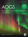
Our work on a “A new bicornuate model of rat uterus transplantation” has been accepted for publication in Acta Obstetricia et Gynecologica Scandinavica.

Uterus transplantation has revolutionized reproductive medicine for women with absolute uterine factor infertility, resulting in more than 40 reported successful live births worldwide to date. Small animal models are pivotal to refine this surgical and immunological challenging procedure aiming to enhance safety for both the mother and the child.
We established a syngeneic bicornuate uterus transplantation model in young female Lewis rats. All surgical procedures were conducted by an experienced and skilled microsurgeon who organized the learning process into multiple structured steps. Animals underwent meticulous preoperative preparation and postoperative care. Transplant success was monitored by sequential biopsies, monitoring graft viability and documenting histological changes long-term. Bicornuate uterus transplantation were successfully established achieving an over 70% graft survival rate with the passage of time. The bicornuate model demonstrated safety and feasibility, yielding outcomes comparable to the unicornuate model in terms of ischemia times and complications. Longitudinal biopsies were well-tolerated, enabling comprehensive monitoring throughout the study. Our novel bicornuate rat uterus transplantation model provides a distinctive opportunity for sequential biopsies at various intervals after transplantation and therefore comprehensive monitoring of graft health, viability, and identification of potential signs of rejection. Furthermore, this model allows for different interventions in each horn for comparative studies without interobserver differences contrary to the established unicornuate model. By closely replicating the clinical setting, this model stands as a valuable tool for ongoing research in the field of uterus transplantation, promoting further innovation and deeper insights into the intricacies of the uterus transplant procedure.

Authors are Dietrich Polenz, Igor Maximilian Sauer, Friederike Martin, Anja Reutzel-Selke, Muhammad Imtiaz Ashraf , Anja Schirmeier , Steffen Lippert, Kirsten Führer, Johann Pratschke, Stefan Günther Tullius, and Simon Moosburner.
Depletion of donor dendritic cells ameliorates immunogenicity of both skin and hind limb transplants
Stacks Image 23686
Our manuscript "Depletion of donor dendritic cells ameliorates immunogenicity of both skin and hind limb transplants" has been accepted for publication in Frontiers in Immunology, section Alloimmunity and Transplantation. Authors are Muhammad Imtiaz Ashraf, Joerg Mengwasser, Anja Reutzel-Selke, Dietrich Polenz, Kirsten Führer, Steffen Lippert, Peter Tang, Edward Michaelis, Rusan Catar, Johann Pratschke, Christian Witzel, Igor M. Sauer, Stefan G. Tullius, and Barbara Kern.

Acute cellular rejection remains a significant obstacle affecting successful outcomes of organ transplantation including vascularized composite tissue allografts (VCA). Donor antigen presenting cells (APC), particularly dendritic cells (DC), orchestrate early alloimmune responses by activating recipient effector T cells. Employing a targeted approach, we investigated the impact of donor-derived conventional DC (cDC) and APC on the immunogenicity of skin and skin-containing VCA grafts, using mouse models of skin and hind limb transplantation.
By post-transplantation day 6, skin grafts demonstrated severe rejections, characterized by predominance of recipient CD4 T cells. In contrast, hind limb grafts showed moderate rejection, primarily infiltrated by CD8 T cells. While donor depletion of cDC and APC reduced frequencies, maturation, and activation of DC in all analysed tissues of skin transplant recipients, reduction in DC activities was only observed in the spleen of hind limb recipients. Donor cDC and APC depletion did not impact all lymphocyte compartments but significantly affected CD8 T cells and activated CD4 T in lymph nodes of skin recipients. Moreover, both donor APC and cDC depletion attenuated the Th17 immune response, evident by significantly reduced Th17 (CD4+IL-17+) cells in the spleen of skin recipients and reduced levels of IL-17E and lymphotoxin-α in the serum samples of both skin and hind limb recipients. In conclusion, our findings underscore the highly immunogenic nature of skin component in VCA. The depletion of donor APC and cDC mitigates the immunogenicity of skin grafts while exerting minimal impact on VCA.
Optimizing environmental enrichment for Sprague Dawley rats: Exemplary insights into the liver proteome
Stacks Image 23706
Considering the expected increase in the elderly population and the growing emphasis on aging-related biomedical research, the demand for aged laboratory animals has surged, challenging established husbandry practices. Our objective was to establish a cost-effective method for environmental enrichment, utilizing the liver as a representative organ to assess metabolic changes in response to differing enrichment levels.
We conducted a six-month study involving 24 male Sprague Dawley rats who were randomly assigned to four environmental enrichment groups. Two groups were housed in standard cages, while the others were placed in modified rabbit cages. Half of the groups received weekly playtime in an enriched rat housing unit. We evaluated hormone levels, playtime behavior, and subjective handling experience. Additionally, liver tissue proteomic analysis was performed.
Initial corticosterone levels and those after 3 and 6 months showed no significant differences. Yet, testosterone levels were lower in the control group by the end of the study (p=0.007). In the liver tissue, we detected 1,871 distinct proteins, with 77% of them being consistent across all groups. In gene ontology analysis, no specific pathways were overexpressed. In semiquantitative analysis, we observed differences in proteins associated in lipid metabolism such as Apolipoprotein A-I and Acyl-CoA 6-desaturase, which were lower in the control group (p= 0.024 and p=0.009). Enriched environments reduced rat distress, large cages eased handling, and conflicts between rats lessened with bi-weekly interactions.

The manuscript "Optimizing environmental enrichment for Sprague Dawley rats: Exemplary insights into the liver proteome" has been accepted for publication in PLOS ONE.
Authors are Nathalie N. Roschke, Karl H. Hillebrandt, Dietrich Polenz, Oliver Klein, Joseph MGV Gassner, Johann Pratschke, Felix Krenzien, Igor M. Sauer, Nathanael Raschzok, and Simon Moosburner.
miRNA as potential biomarkers after liver transplantation: A systematic review
Stacks Image 23730
The publication "miRNA as potential biomarkers after liver transplantation: A systematic review" is now available online in Transplantation Reviews. Authors are Pia F. Koch, Kristina Ludwig, Felix Krenzien, Karl H. Hillebrandt, Wenzel Schöning, Johann Pratschke, Nathanael Raschzok, Igor M. Sauer, and Simon Moosburner.

Early and accurate diagnosis of acute cellular rejection (ACR) and graft complications after liver transplantation is crucial for timely intervention and improved patient outcomes, but their diagnosis rely currently on invasive biopsy sampling, thus prompting the search for non-invasive Biomarkers. MicroRNA (miRNA) have emerged as promising biomarkers in various pathological conditions, and their potential utility in diagnosing acute cellular rejection after liver transplantation has gained significant interest.

This systematic review analyzes studies exploring miRNA as biomarkers for ACR and graft dysfunction in liver transplantation (PROSPERO ID CRD42023465278). The Cochrane Collaboration tool for assessing risk of bias was employed. Population data, identified miRNA and their dynamic regulation, as well as event prediction were compared. Data extraction and quality assessment were performed independently by two reviewers.

The results highlight the potential of miRNA as specific, non-invasive biomarkers for ACR and graft dysfunction following liver transplantation. However, further research is needed to validate these findings and establish standardized diagnostic panels to incorporate them into clinical practice and explore miRNA-based therapies in the future.
Rise of tissue- and species-specific 3D bioprinting based on decellularized extracellular matrix-derived bioinks and bioresins
Stacks Image 23761
The publication "Rise of tissue- and species-specific 3D bioprinting based on decellularized extracellular matrix-derived bioinks and bioresins" is now available online in Biomaterials and Biosystems. Authors are Laura Elomaa, Ahed Almalla, Eriselda Keshi, Karl H. Hillebrandt, Igor M. Sauer, and Marie Weinhart.

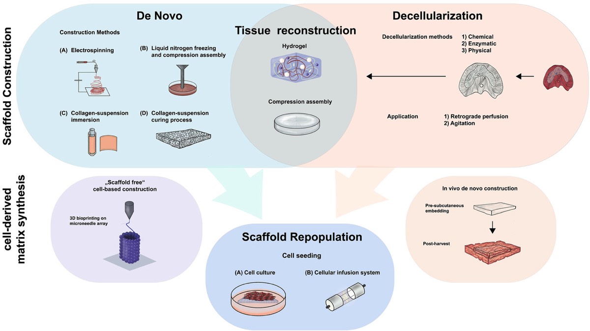
Thanks to its natural complexity and functionality, decellularized extracellular matrix (dECM) serves as an excellent foundation for creating highly cell-compatible bioinks and bioresins. This enables the bioprinted cells to thrive in an environment that closely mimics their native ECM composition and offers customizable biomechanical properties. To formulate dECM bioinks and bioresins, one must first pulverize and/or solubilize the dECM into non-crosslinked fragments, which can then be chemically modified as needed. In bioprinting, the solubilized dECM-derived material is typically deposited and/or crosslinked in a layer-by-layer fashion to build 3D hydrogel structures. Since the introduction of the first liver-derived dECM-based bioinks, a wide variety of decellularized tissue have been employed in bioprinting, including kidney, heart, cartilage, and adipose tissue among others. This review aims to summarize the critical steps involved in tissue-derived dECM bioprinting, starting from the decellularization of the ECM to the standardized formulation of bioinks and bioresins, ultimately leading to the reproducible bioprinting of tissue constructs.
Moderate LMWH anticoagulation improves success rate of hind limb allotransplantation in mice
Stacks Image 23773
The publication "Moderate LMWH Anticoagulation Improves Success Rate of Hind Limb Allotransplantation in Mice" is now available online in Plastic & Reconstructive Surgery-Global Open. Authors are B. Kern, M.-I. Ashraf, A. Reutzel-Selke, J. Mengwasser, D. Polenz, E. Michaels, J. Pratschke, S.G. Tullius, Ch. Witzel, and I.M. Sauer.

The mouse hind limb model represents a powerful research tool in vascularized composite tissue allotransplantation, but its applicability is limited due to poor graft survival (62%–83%). Vascular thrombosis and massive hemorrhage are the major causes for these drop-outs. We hypothesize that because of better anticoagulation effect and lower risk of thrombocytopenia, application of low molecular weight heparin (LMWH) will minimize vascular complications and enhance graft and animal survival.

Fifty allogeneic hind limb transplantations were performed (C57BL/6 to DBA/2 mice) using five different anticoagulation protocols. Bleeding and thromboembolic events were recorded macroscopically by postoperative hemorrhage and livid discoloration of the graft, respectively. Graft perfusion and survival were monitored daily by capillary-refill-time of graft toes within 2–3 seconds. Vascular congestion and tissue necrosis were examined by histological evaluation of hematoxylin-eosin-stained tissue sections.

All transplantations were technically successful. Increase in thromboembolic events and a concomitant decrease in bleeding events were observed with the decreasing concentration of heparin in the perfusion solution. Although treatment of donor and recipient with low dose of LMWH could not reduce thromboembolic events, moderate dose effectively reduced these events. Compared with the poor outcome of graft perfusion with heparin alone, additional treatment of donor and recipient with low dose of LMWH improved graft and animal survival by 18%. Interestingly, animals treated with moderate dose of LMWH demonstrated 100% graft and animal survival.
Treatment of donor and recipient mice with a moderate dose of LMWH prevents vascular complications and improves the outcome of murine hind limb transplants.
 Page 1 / 1 
Stacks Image 27655
Our manuscript "Depletion of donor dendritic cells ameliorates immunogenicity of both skin and hind limb transplants" has been accepted for publication in Frontiers in Immunology, section Alloimmunity and Transplantation. Authors are Muhammad Imtiaz Ashraf, Joerg Mengwasser, Anja Reutzel-Selke, Dietrich Polenz, Kirsten Führer, Steffen Lippert, Peter Tang, Edward Michaelis, Rusan Catar, Johann Pratschke, Christian Witzel, Igor M. Sauer, Stefan G. Tullius, and Barbara Kern.

Acute cellular rejection remains a significant obstacle affecting successful outcomes of organ transplantation including vascularized composite tissue allografts (VCA). Donor antigen presenting cells (APC), particularly dendritic cells (DC), orchestrate early alloimmune responses by activating recipient effector T cells. Employing a targeted approach, we investigated the impact of donor-derived conventional DC (cDC) and APC on the immunogenicity of skin and skin-containing VCA grafts, using mouse models of skin and hind limb transplantation.
By post-transplantation day 6, skin grafts demonstrated severe rejections, characterized by predominance of recipient CD4 T cells. In contrast, hind limb grafts showed moderate rejection, primarily infiltrated by CD8 T cells. While donor depletion of cDC and APC reduced frequencies, maturation, and activation of DC in all analysed tissues of skin transplant recipients, reduction in DC activities was only observed in the spleen of hind limb recipients. Donor cDC and APC depletion did not impact all lymphocyte compartments but significantly affected CD8 T cells and activated CD4 T in lymph nodes of skin recipients. Moreover, both donor APC and cDC depletion attenuated the Th17 immune response, evident by significantly reduced Th17 (CD4+IL-17+) cells in the spleen of skin recipients and reduced levels of IL-17E and lymphotoxin-α in the serum samples of both skin and hind limb recipients. In conclusion, our findings underscore the highly immunogenic nature of skin component in VCA. The depletion of donor APC and cDC mitigates the immunogenicity of skin grafts while exerting minimal impact on VCA.

Archive


Categories

Year

This website or its third-party tools use cookies, which are necessary to its functioning and required to achieve the purpose illustrated in the Disclaimer. By closing this banner, scrolling this page, clicking a link or continuing to browse otherwise, you agree to the use of cookies.