News

Magnetic resonance elastography quantification of decellularized liver tissue
Stacks Image 24242
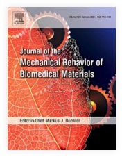
"Magnetic resonance elastography quantification of the solid-to-fluid transition of liver tissue due to decellularization" was published in the latest issue of the Journal of the Mechanical Behavior of Biomedical Materials.

Maintenance of tissue extracellular matrix (ECM) and its biomechanical properties for tissue engineering is one of the substantial challenges in the field of decellularization and recellularization. Preservation of the organ-specific biomatrix is crucial for successful recellularization to support cell survival, proliferation, and functionality. However, understanding ECM properties with and without its inhabiting cells as well as the transition between the two states lacks appropriate test methods capable of quantifying bulk viscoelastic parameters in soft tissues.
We used compact magnetic resonance elastography (MRE) with 400, 500, and 600 Hz driving frequency to investigate rat liver specimens for quantification of viscoelastic property changes resulting from decellularization. Tissue structures in native and decellularized livers were characterized by collagen and elastin quantification, histological analysis, and scanning electron microscopy.
Decellularization did not affect the integrity of microanatomy and structural composition of liver ECM but was found to be associated with increases in the relative amounts of collagen by 83-fold (37.4 ± 17.5 vs. 0.5 ± 0.01 μg/mg, p = 0.0002) and elastin by approx. 3-fold (404.1 ± 139.6 vs. 151.0 ± 132.3 μg/mg, p = 0.0046). Decellularization reduced storage modulus by approx. 9-fold (from 4.9 ± 0.8 kPa to 0.5 ± 0.5 kPa, p < 0.0001) and loss modulus by approx. 7-fold (3.6 kPa to 0.5 kPa, p < 0.0001), indicating a marked loss of global tissue rigidity as well as a property shift from solid towards more fluid tissue behavior (p = 0.0097).
Our results suggest that the rigidity of liver tissue is largely determined by cellular components, which are replaced by fluid-filled spaces when cells are removed. This leads to an overall increase in tissue fluidity and a viscous drag within the relatively sparse remaining ECM. Compact MRE is an excellent tool for quantifying the mechanical properties of decellularized biological tissue and a promising candidate for useful applications in tissue engineering.

Authors are Hannah Everwien, Angela Ariza de Schellenberger, Nils Haep, Heiko Tzschätzsch, Johann Pratschke, Igor M. Sauer, Jürgen Braun, Karl H. Hillebrandt and Ingolf Sack.

J Mech Behav Biomed Mater. 2020 Apr;104:103640. doi: 10.1016/j.jmbbm.2020.103640. Epub 2020 Jan 14.
Back
Stacks Image 27655
Our manuscript "Depletion of donor dendritic cells ameliorates immunogenicity of both skin and hind limb transplants" has been accepted for publication in Frontiers in Immunology, section Alloimmunity and Transplantation. Authors are Muhammad Imtiaz Ashraf, Joerg Mengwasser, Anja Reutzel-Selke, Dietrich Polenz, Kirsten Führer, Steffen Lippert, Peter Tang, Edward Michaelis, Rusan Catar, Johann Pratschke, Christian Witzel, Igor M. Sauer, Stefan G. Tullius, and Barbara Kern.

Acute cellular rejection remains a significant obstacle affecting successful outcomes of organ transplantation including vascularized composite tissue allografts (VCA). Donor antigen presenting cells (APC), particularly dendritic cells (DC), orchestrate early alloimmune responses by activating recipient effector T cells. Employing a targeted approach, we investigated the impact of donor-derived conventional DC (cDC) and APC on the immunogenicity of skin and skin-containing VCA grafts, using mouse models of skin and hind limb transplantation.
By post-transplantation day 6, skin grafts demonstrated severe rejections, characterized by predominance of recipient CD4 T cells. In contrast, hind limb grafts showed moderate rejection, primarily infiltrated by CD8 T cells. While donor depletion of cDC and APC reduced frequencies, maturation, and activation of DC in all analysed tissues of skin transplant recipients, reduction in DC activities was only observed in the spleen of hind limb recipients. Donor cDC and APC depletion did not impact all lymphocyte compartments but significantly affected CD8 T cells and activated CD4 T in lymph nodes of skin recipients. Moreover, both donor APC and cDC depletion attenuated the Th17 immune response, evident by significantly reduced Th17 (CD4+IL-17+) cells in the spleen of skin recipients and reduced levels of IL-17E and lymphotoxin-α in the serum samples of both skin and hind limb recipients. In conclusion, our findings underscore the highly immunogenic nature of skin component in VCA. The depletion of donor APC and cDC mitigates the immunogenicity of skin grafts while exerting minimal impact on VCA.

Archive


Categories

Year

This website or its third-party tools use cookies, which are necessary to its functioning and required to achieve the purpose illustrated in the Disclaimer. By closing this banner, scrolling this page, clicking a link or continuing to browse otherwise, you agree to the use of cookies.