News

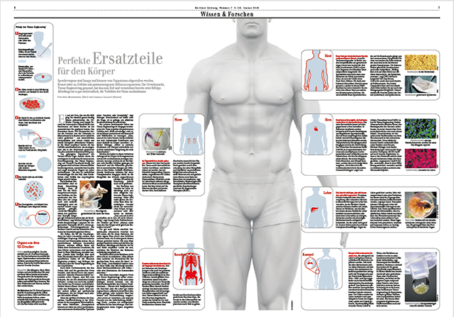
HCC: miRNA profiles of the tumor-surrounding tissue
Stacks Image 24778
The presence of hepatocellular carcinoma (HCC) is a significant complication of cirrhosis because it changes the prognosis and the treatment of the patients. By now, contrast-enhanced CT and MR scans are the most reliable tools for the diagnosis of HCC; however, in some cases, a biopsy of the tumor is necessary for the final diagnosis. The aim of the study was to develop a diagnostic tool using the microRNA (miRNA) profiles of the tissue surrounding the HCC tumor combined with clinical parameters in statistical models. At a transplantation setting, 32 patients with HCC and cirrhosis (B) were compared to 22 patients suffering from cirrhosis only (A). The diagnosis and exclusion of HCC was confirmed following the histopathological examination of the explanted liver.

The HCC patients were significantly older than the patients with cirrhosis only (B: 60.6 and A: 49.9, p<0.001) and showed higher levels of ALT (A: 0.76μkat/l, B: 1.02μkat/, p=0.006) and AFP (A: 5.8ng/ml, B: 70.3ng/ml, p<0.001), whereas the bilirubin levels were higher in the cirrhosis only group (p=0.002). Using age (cut-off 50.23years) and AFP (cut-off 4.2ng/ml) thresholds, the levels of expression of miR-1285-3p and miR-943 differentiated between the patients with HCC and cirrhosis from those with cirrhosis only with an accuracy of 96.3%. This is the first report about the use of stepwise penalized logistic regression and decision tree analyses of miRNA expressions in the tumor-surrounding tissue combined with clinical parameters for the diagnosis of HCC.

Authors are Mehmet Haluk Morgul, Sergej Klunk, Zografia Anastasiadou, Ulrich Gauger, Corinna Dietel, Anja Reutzel-Selke, Philipp Felgendref, Hans-Michael Hau, Hans-Michael Tautenhahn, Rosa Bianca Schmuck, Nathanael Raschzok, Igor Maximillian Sauer, and Michael Bartels. Exp Mol Pathol. 2016 Aug 20;101(2):165-171. doi: 10.1016/j.yexmp.2016.07.014. [Epub ahead of print]
Read More
Bile: miRNA Pattern post OLT
Stacks Image 24788
BIOMARKERS accepted our latest paper on "Bile: miRNA Pattern and Protein Based Biomarkers May Predict Acute Cellular Rejection after Liver Transplantation" for publication. Authors are Rosa Bianca Schmuck, Anja Reutzel-Selke, Nathanael Raschzok, Mehmet Haluk Morgul, Benjamin Struecker, Steffen Lippert, Cynthia de Carvalho Fischer, Moritz Schmelzle, Sabine Boas-Knoop, Marcus Bahra, Andreas Pascher, Johann Pratschke, and Igor M. Sauer.

Bile rather than blood depicts the local inflammation in the liver and may improve prediction and diagnosis of acute cellular rejection (ACR) after liver transplantation (OLT). Secretome and miRNAs were analyzed during the first two weeks and on clinical suspicion of ACR in the bile of 45 OLT recipients. Levels of CD44, CXCL9, miR-122, miR-133a, miR-148a and miR-194 were significantly higher in bile of patients who developed ACR within the first 6 months after OLT and during ACR. Analysis of secretome and miRNA in bile could further our understanding of the local inflammatory process during rejection.
Read More
Hepatocyte isolation after laparoscopic liver resection
Stacks Image 24796
Tissue Engineering, Part C: Methods accepted our paper entitled "Hepatocyte isolation after laparoscopic liver resection" for publication. Authors are Horner R*, Kluge M*, Gassner J, Nösser M, Major RD, Reutzel-Selke A, Leder AK, Struecker B, Morgul MH, Pratschke J, Sauer IM, Raschzok N (*contributed equally).

Liver tissue obtained from partial hepatectomy is a common source for isolation of primary human hepatocytes. Until now, liver resections were most commonly performed by conventional open surgery. Although the laparoscopic approach is currently emerging in liver surgery, data on the outcome of hepatocyte isolation from laparoscopically resected liver tissue is not available. A total of 22 hepatocyte isolations were performed using the two-step collagenase perfusion technique from October 2015 until March 2016. Liver tissue was obtained from n=15 open liver resections (OLR) and n=7 laparoscopic liver resections (LLR). Isolation parameters (cell yield, viability, percoll survival) were assessed and hepatocyte function (plating efficiency, urea, albumin, and aspartate aminotransferase) was measured over a culture period of 6 days (OLR: n=13; LLR: n=3). Total cell yield (OLR: 36.81 ± 6.77 x10⁶ cells/g vs. LLR 16.84 ± 10.66 x10⁶ cells/g, p=0.0318) as well as viable yield (OLR 31.70 ± 6.05 x10⁶ cells/g vs. LLL 14.70 ± 9.89 x10⁶ cells/g, p=0.0260) were significantly higher in the OLR group. Subgroup analysis revealed that the worse outcome of isolation of laparoscopically resected liver tissue was associated with right-lateral laparoscopic liver resections, while hepatocyte isolation from left-lateral laparoscopic liver resections was as effective as from open surgery. Hepatocyte function did not differ between hepatocytes from openly resected versus left-lateral laparoscopically resected liver tissue. We here present the first data on hepatocyte isolation from laparoscopic liver surgery. While the overall outcome is worse compared to open surgery, our data suggest that liver tissue from laparoscopic resection of the left lobe is an excellent source for primary human hepatocytes.
Read More
SPARK Berlin supports Fikatas Knot
Stacks Image 24815
The Berlin Institute of Health (BIH) and Stiftung Charité teamed up with Stanford University School of Medicine to initiate SPARK Berlin.

We are very pleased to announce that Dr. Panagiotis Fikatas' project “Device for ready-prepared surgical knots” was selected for both, funding and mentorship. 

SPARK was created to overcome the hurdles associated with translating academic discoveries into therapeutics and diagnostics that address unmet medical needs.  The SPARK mission is to help academics overcome the obstacles involved in moving their early discoveries from bench to bedside, to educate faculty, postdoctoral fellows and graduate students on the translational research process and path to clinical application, so that development of promising discoveries becomes second nature to our institution, and to develop more cost-effective and innovative approaches to drug development .

Congratulations!
Read More
International Medical Tournament 2016
Stacks Image 24834
Karl Hillebrandt and his team won this year's International Medical Tournament (English League) in Novosibirsk, Russia!
Congratulations!

The International Medical Tournament is a team competition for future doctors, which provides unique professional experience such as solving complex diagnostic cases from real clinical practice without any risks, presentation and discussion of solutions among students and experts. The main goal of the competition is to help medical students gather and develop lacking skills for future medical practice, critical thinking in diagnostic process, abilities of teamwork, presentation and rhetorical skills.  
Read More
NeoHybrid liver graft – proof of concept
Stacks Image 24842
Cells Tissues Organs accepted our latest paper on "Allogeneic liver transplantation and subsequent syngeneic hepatocyte transplantation in a rat model – proof of concept for in vivo tissue engineering" for publication.

Authors are Susanne Rohn, Jan Schroeder, Henriette Riedel, Dietrich Polenz, Katarina Stanko, Anja Reutzel-Selke, Peter Tang, Lydia Brusendorf, Nathanael Raschzok, Peter Neuhaus, Johann Pratschke, Birgit Sawitzki, Igor M. Sauer, and Martina T. Mogl.

Aim of the project was the evaluation of a new concept for in vivo tissue engineering using autologous primary human hepatocytes and hepatic progenitor cells isolated from diseased livers explanted during orthotopic liver transplantation (LTx). Cells will be isolated and infused into the spleen for repopulation of the allogeneic liver graft. The latter is serving as biological matrix for the engraftment of autologous cells. Once these cells have engrafted, it is assumed that autologous cells will repopulate the allogeneic liver, since they should have a selective advantage due to their autologous origin. It is postulated that this will lead to a neo-hybrid liver graft, reducing immunogenicity and inducing immunoregulation thus minimizing the need for extensive immunosuppression and eventually inducing operational tolerance. 

We therefore developed a new rat model for combined liver and liver cell transplantation under stable immunosuppression. Immunohistochemistry demonstrated the engraftment of transplanted cells, as confirmed by fluorescence in-situ hybridization, showing repopulation of the liver graft with 15.6 % male cells (± 1.8 SEM) at day 90. The quantitative PCR revealed 14.15 % (mean ± 5.09 SEM) male DNA at day 90. Engraftment of transplanted autologous cells after combined liver and cell transplantation was achieved for up to 90 days under immunosuppression. Immunohistochemistry indicated cell proliferation, and the fluorescence in-situ hybridization results were partly confirmed by quantitative RT-PCR. This new protocol in rats appears feasible to address long-term function and eventually induction of operational tolerance in the future.
Read More
LTx – microRNA signatures in peripheral blood ?
Stacks Image 24852
BIOMARKERS accepted our latest paper on „microRNA signatures in peripheral blood fail to detect acute cellular rejection after liver transplantation“ for publication. Authors are N. Raschzok, A. Reutzel-Selke, R. Schmuck, L. Tannus, M. Morgul, C. Dietel, A. Leder, B. Struecker, S. Lippert, H. Sallmon, M. Schmelzle, M. Bartels, S. Jonas, J. Pratschke, and I.M. Sauer.

We investigated whether microRNA signatures in whole blood samples are associated with acute cellular rejection (ACR) after liver transplantation. Blood samples were collected using Paxgene technology and analyzed by microarrays and qRT-PCR. microRNA signatures failed to distinguish between 19 patients with ACR and 16 controls. Let-7b-5p and let-7c were up-regulated in a subgroup of patients with ACR during the 6th and 7th postoperative day but failed in an independent validation of 20 patients. microRNA signatures in whole blood processed by Paxgene technology are not suited for detection of ACR after liver transplantation.
Read More
Single Pass Albumin Dialysis – Dose finding study
Stacks Image 24862
Artificial Organs accepted our paper „Single Pass Albumin Dialysis (SPAD) – A dose finding study to define optimal albumin concentration and dialysate flow“ for publication.  Authors are R.B. Schmuck, G.-H. Nawrot, P. Fikatas, A. Reutzel-Selke, J. Pratschke, and I.M. Sauer.

Aim of these studies was to define the optimal conditions for SPAD in a standardized experimental set-up. Albumin concentration was adjusted to either 1%, 2%, 3%, or 4%, while the flow rate of the dialysate was kept constant at a speed of 700 ml/h. The flow rate of the dialysate was altered between 350, 500, 700, and 1000 ml/h, whereas the albumin concentration was continuously kept at 3%. 

This study revealed that the detoxification of albumin bound substances could be improved by increasing the concentration of albumin in the dialysate with an optimum at 3%. A further increase of the albumin concentration to 4% did not lead to a significant increase in detoxification. Furthermore, we observed a gradual increase of the detoxification efficiency for albumin bound substances, from 350 ml/h to 700 ml/h (for bilirubin) or 1000 ml/h (for bile acids) of dialysate flow. Water-soluble toxins (ammonia, creatinine, urea, uric acid) were removed almost completely, regardless of albumin concentration or flow rate. 
Read More
ESOT | YPT – Interview with Karl Hillebrandt
Stacks Image 24884
Being the winner of the Rising Stars Video Session organised by the YPT Committee at the ESOT2015 Brussels Congress Karl Hillebrandt gave an interview for the ESOT | YPT webpage.

His abstract "Optimized decellularization of rat livers byarterial and portal venous perfusion underoscillating pressure conditions" and the accompanying video were the most voted at the Rising Stars Session, where the audience voted live for the best video abstract. You can read Karl's abstract in the special ESOT2015 issue of Transplant International.
Read More
Implantation of a Neo Bile Duct in domestic pigs
Stacks Image 24901
European Surgical Research accepted our latest paper entitled "Implantation of a tissue engineered Neo Bile Duct in domestic pigs" for publication. Authors are B. Struecker, K. Hillebrandt, N. Raschzok, K. Jöhrens, A. Butter, P. Tang, A. Andreou, H. Napierala, D. Polenz, A. Reutzel-Selke, T. Denecke, J. Pratschke, and I.M Sauer.

Extrahepatic bile duct injuries remain severe complications during cholecystectomy and often require reconstruction by bilioenteric anastomosis (i.e. hepatico-jejunostomy), which comes along with further long-term complications (e.g. recurring ascending cholangitis, secondary biliary cirrhosis). Furthermore, in case of inherent extrahepatic biliary atresia or during liver transplant artificial or engineered bile ducts could enable novel surgical strategies without the need for hepatico-jejunostomy. We present data on the implantation of in vitro generated Neo Bile Ducts in five domestic pigs. Neo Bile Ducts were engineered through decellularization of allogeneic blood vessels and recellularization with autologous cholangiocytes.On postoperative days 0, 1, 7 and 14 blood samples were taken and analyzed (AST, ALT, Bilirubin, Alkaline Phosphatase, Creatinine and Leukocytes). An magnetic resonance cholangiography was performed on postoperative day 14 with one pig. 14 days after implantation pigs were sacrificed and bile ducts were explanted. All pigs survived the complete study period without severe complications. None of the pigs showed signs of biliary leakage or peritonitis. Neo Bile Ducts were infiltrated by neutrophils and neo-angiogenesis was observed around and into the implanted tissue. Whether the presented technique enables the long-term replacement of native bile ducts has to be further evaluated.
Read More
Human hepatocyte isolation – new paper
Stacks Image 24911
Tissue Engineering, Part C: Methods accepted our paper „Human hepatocyte isolation: Does portal vein embolization affect the outcome?“ Authors are Martin Kluge, Anja Reutzel-Selke, Hendrik Napierala, Karl H. Hillebrandt, Rebeka D. Major, Benjamin Struecker, Annekatrin Leder, Jeffrey Siefert, Peter Tang, Steffen Lippert, Daniel Seehofer, Johann Pratschke, Igor M. Sauer und Nathanael Raschzok.

Primary human hepatocytes are widely used for basic research, pharmaceutical testing, and therapeutic concepts in regenerative medicine. Human hepatocytes can be isolated from resected liver tissue. Preoperative portal vein embolization (PVE) is increasingly used to decrease the risk of delayed postoperative liver regeneration by induction of selective hypertrophy of the future remnant liver tissue. The aim of this study was to investigate the effect of PVE on the outcome of hepatocyte isolation. Primary human hepatocytes were isolated from liver tissue obtained from partial hepatectomies (n=190) using the two-step collagenase perfusion technique followed by Percoll purification. Of these hepatectomies, 27 isolations (14.2%) were performed using liver tissue obtained from patients undergoing PVE prior to surgery. All isolations were characterized using parameters that had been described in the literature as relevant for the outcome of hepatocyte isolation. The PVE and non-PVE groups were similar in regard to donor parameters (sex, age, indication for surgery), isolation parameters (liver weight, cold ischemic time), and the quality of the liver tissue. The mean initial viable cell yield did not differ between the PVE and non-PVE groups (10.16±2.03x10⁶ cells/g vs. 9.70±0.73 x10⁶ cells/g, p=0.499). The initial viability was slightly better in the PVE-group (77.8 ±2.03% vs. 74.4 ±1.06%). The mean viable cell yield (p=0.819) and the mean viability (p=0.141) after Percoll purification did not differ between the groups. PVE had no effect on enzyme leakage and metabolic activity of cultured hepatocytes.  Although PVE leads to drastic metabolic alterations and changes in hepatic blood flow, embolized liver tissue is a suitable source for the isolation of primary human hepatocytes and is equivalent to untreated liver tissue in regard to cell yield and viability.
Read More
Dr. Jan Schröder
Today, Jan Schröder successfully defended his thesis (summa cum laude) entitled "Vergleichende in vivo und in vitro Analysen im Rahmen der Etablierung der kombinierten Leber- und Leberzelltransplantation im Rattenmodell" !

Congratulations !
Read More
Karl Hillebrandt – YPT Rising Star
Stacks Image 24925

Karl Hillebrandt won this year's YPT Rising Stars Video Session Award. He presented the studies on „Optimized decellularization of rat livers by arterial and portal venous perfusion under oscillating pressure conditions“ during the 17th congress of the European Society for Organ Transplantation (ESOT) 2015 in Brüssel. Young Professionals in Transplantation (YPT) is a forum for junior professionals throughout Europe working in the field of transplantation.

Read More
The Morning After
Referring to our paper „CD44 and CXCL9 serum protein levels predict the risk of clinically significant allograft rejection after liver transplantation“ Geoffrey W. McCaughan, Patrick Bertolino and David G. Bowen wrote an interesting editorial entitled „Could The Morning After liver transplant be immunologically interesting?“ 

They conclude, „that our study urges us to study the immune system response in liver allograft recipients during the very early phases after liver transplantation and to explore how events in immune organs and the allograft are reflected within the serum. Whether the patterns observed truly represent early detection of ACR versus tolerance, or a combination of both, requires further study and experimentation, including the identification of the cellular sources of these and other potential markers of immune outcome. It seems that despite significant levels of immunosuppressive drugs, immune activation and engagement occurs very early after human liver transplant, within the first 24 hours, in a manner that may have similarities with experimental animal models. Thus, the morning after effect could be an exciting window to longer-term immune outcomes, rather than just being preoccupied with observing important routine outcomes and detecting early complications.“
Read More
Publication in Jove
Procedure for Decellularization of Rat Livers in an Oscillating-pressure Perfusion Device“ is available in J Vis Exp. 2015 Aug 10;(102). doi: 10.3791/53029 – accessible via the JOVE servers. 

Authors are K. Hillebrandt, D. Polenz, A. Butter, P. Tang, A. Reutzel-Selke, A. Andreou, H. Napierala, N. Raschzok, J. Pratschke, I.M. Sauer, and B. Struecker . The presented techniques for liver harvesting, cannulation and perfusion using our proprietary device enable sophisticated perfusion set-ups to improve decellularization and recellularization experiments in rat livers.
Read More
microRNAs in liver tissue engineering
Stacks Image 24955
Our paper "microRNAs in liver tissue engineering - New promises for failing organs"was accepted for publication in Advanced Drug Delivery Reviews (IF: 15.038). Authors are Nathanael Raschzok, Hannes Sallmon, Johann Pratschke and Igor M. Sauer.

miRNA-based technologies provide attractive tools for several liver tissue engineering approaches. Herein, we review the current state of miRNA applications in liver tissue engineering. Several miRNAs have been implicated in hepatic disease and proper hepatocyte function. However, the clinical translation of these findings into tissue engineering has just begun. miRNAs have been successfully used to induce proliferation of mature hepatocytes and improve the differentiation of hepatic precursor cells. Nonetheless, miRNA-based approaches beyond cell generation have not yet entered preclinical or clinical investigations. Moreover, miRNA-based concepts for the biliary tree have yet to be developed. Further research on miRNA based modifications, however, holds the promise of enabling significant improvements to liver tissue engineering approaches due to their ability to regulate and fine-tune all biological processes relevant to hepatic tissue engineering, such as proliferation, differentiation, growth, and cell function.
Read More
CD44 and CXCL9 predicting rejection after LTx
Stacks Image 24965
Based on a fruitful collaboration with the department of Visceral, Transplantation, Thoracic, and Vascular Surgery at the University of Leipzig our paper on „CD44 and CXCL9 serum protein levels predict the risk of clinically significant allograft rejection after liver transplantation“ has been accepted for publication in Liver Transplantation. 

Authors are Nathanael Raschzok, Anja Reutzel-Selke, Rosa Bianca Schmuck, Mehmet Haluk Morgul, Ulrich Gauger, Kukuh Aji Prabowo, Laura-Marie Tannus, Annekatrin Leder, Benjamin Struecker, Sabine Boas-Knoop, Michael Bartels, Sven Jonas, Christian Lojewski, Gero Puhl, Daniel Seehofer, Marcus Bahra, Andreas Pascher, Johann Pratschke, and Igor Maximilian Sauer.

The diagnosis of acute cellular rejection (ACR) after liver transplantation is based on histological analysis of biopsies because non-invasive biomarkers for allograft rejection are not yet established for clinical routines. CD31, CD44, and CXCL9 have previously been described as biomarkers for cross-organ allograft rejection. Here, we assessed the predictive and diagnostic value of these proteins as serum biomarkers for clinically significant ACR in the first six months after liver transplantation in a prospective study. The protein levels were measured in 94 patients immediately prior to transplantation, at postoperative days (POD) 1, 3, 7, and 14, and when biopsies were performed during episodes of biochemical graft dysfunction. Our results suggest that CD44 and CXCL9 may serve as predictive biomarkers to identify liver allograft recipients at risk for clinically significant ACR.
Read More
Cover – march issue of Tissue Engineering
Stacks Image 24970

One of the figures of our paper „Porcine liver decellularization under oscillating pressure conditions – A technical refinement to improve the homogeneity of the decellularization process“ made it to the cover of the march issue of Tissue Engineering, Part C : Methods.

Congratulations to Dietrich Polenz, who made the corrosion cast of a decellularized pig liver matrix: red, hepatic artery; blue, portal vein; yellow, bile duct and gallbladder.

Read More
Particles for microRNA-targeted manipulation
Biomaterials accepted our paper on „Micron-sized iron oxide-containing particles for microRNA-targeted manipulation and MRI-based tracking of transplanted cells“. Authors are Annekatrin Leder, Nathanael Raschzok, Christian Schmidt, Duygu Arabacioglu, Antje Butter, Susanne Kolano, Luisa S. de Sousa Lisboa, Wiebke Werner, Dietrich Polenz, Anja Reutzel-Selke, Johann Pratschke, and Igor M. Sauer.

Particle-based delivery systems for therapeutic manipulation and tracking of transplanted cells by magnetic resonance imaging (MRI) are commonly based on nanometer-sized superparamagnetic iron oxide particles (SPIOs). Here, we present a proof of concept for multifunctional, silica based micron-sized iron oxide-containing particles (sMPIO) that combine fluorescence imaging, MRI tracking, and on-the-spot targeting of specific microRNAs on a particle surface for therapeutic manipulation by RNA interference. Antisense locked nucle-LNA) were covalently bound to the surface of silica-based, DAPI-integrated, micron-sized iron oxide particles (sMPIO--LNA). In vitro studies using primary human hepatocytes showed rapid particle uptake (4 hours) that was accompanied by significant depletion of the targeted microRNA Let7g (80%), up- regulation of the target proteins Cyclin D1 and c-Myc, and specific proteome changes. sMPIO--LNA- labeled cells were successfully detected by fluorescence imaging and could be visualized by MRI after intrasplenic transplantation in rats. This new theranostic particle provides a promising tool for cell transplantation where cellular imaging and microRNA-based manipulation is needed.

Biomaterials is the leading journal in its field. Impact factors released by ISI in July 2014 showed Biomaterials with an impact factor of 8.312!
Read More
Decellularization of pancreata – EPITA Award
Stacks Image 24991
Ben Strücker presented our latest results on DECELLULARIZATION OF WHOLE RAT PANCREATA – EVALUATION OF THREE DIFFERENT PERFUSION ROUTES at the 5th EPITA Winter Symposium in Innsbruck from the 25th to the 27th of January 2015. He received the AIDPIT&EPITA Award for the best oral presentation. Congratulations!

Ben Strücker presented three effective protocols for rat pancreas perfusion decellularization, evaluating different perfusion routes. In contrast to liver decellularization the perfusion route seems to have no major impact on decellularization results. The dPECMs could serve for cellular repopulation with islets from a different (xenogene) origin to generate functional, transplantable endocrine pancreata in vitro.
Read More
Rebeka Major: BIH Promotionsstipendium
Rebeka Major successfully applied for the BIH-Promotionsstipendium grant. Her work will focus in the role of the INDY („I’m Not Dead Yet“) gene in liver regeneration – a project in cooperation with Prof. A. Birkenfeld, Universitätsklinikum Carl Gustav Carus in Dresden.
In the Berlin Institute of Health (BIH), the Max Delbrück Center for Molecular Medicine (MDC) and the Charité - Universitätsmedizin Berlin have joined forces. The core idea is to combine translational research with an overarching systems medicine approach to bridge the gap between basic research and clinical application.
Read More
Presentations at DTG 2014
Stacks Image 25015
Nathanael Raschzok presented his poster „Protein biomarkers for diagnosis and prediction of acute cellular rejection after liver transplantation“, Benjamin Strücker his work entitled „Oszillierende Druckschwankung verbessern signifikant die Ratten- und Schweineleber Dezellularisierung“ and Rosa Schmuck her posters „Risk of postoperative infections after LTX rises with MELD score“ and „Protein biomarkers in bile as a diagnostic tool after liver transplantation“.
Read More
Ben Strücker: Charité Clinical Scientist
Stacks Image 25025
Dr. med Benjamin Strücker successfully applied for the Charité Clinical Scientist 2015 program.
His project is entitled „Humanized Porcine Liver““. Clinical mentor is Prof. Dr. Johann Pratschke, scientific mentors is Priv.-Doz. Dr. med Igor M. Sauer.

The program is supported by Stiftung Charité which was endowed by the entrepreneur Johanna Quandt in order to promote biomedical "knowledge entrepreneurs" that is, change makers in biomedicine at the Charité. The goal of this program is to develop new career paths in clinical specialist medical training. The focus of the training program "Clinical Scientist" is translational research ("bench-to-bedside") which will be realized by a reduction in clinical routine and an improved curriculum with defined goals.
Read More
Sham Laparotomy and microRNA Expression in Rats
Stacks Image 25035
BMC Research Notes accepted our latest paper on „Independent Effects of Sham Laparotomy and Anesthesia on Hepatic microRNA Expression in Rats“ for publication.

Studies on liver regeneration following partial hepatectomy (PH) have identified several microRNAs (miRNAs) that show a regulated expression pattern. These studies involve major surgery to access the liver, which is known to have intrinsic effects on hepatic gene expression and may also affect miRNA screening results. We performed two-third PH or sham laparotomy (SL) in Wistar rats to investigate the effect of both procedures on miRNA expression in liver tissue and corresponding plasma samples by microarray and qRT-PCR analyses. As control groups, non-treated rats and rats undergoing anesthesia only were used. We found that 49 out of 323 miRNAs (15%) were significantly deregulated after PH in liver tissue 12 to 48 hours postoperatively (>20% change), while 45 miRNAs (14%) were deregulated following SL. Out of these miRNAs, 10 miRNAs were similarly deregulated after PH and SL, while one miRNA showed opposite regulation. In plasma, miRNA upregulation was observed for miR-133a and miR-133b following PH and SL, whereas miR-100 and miR-466c were similarly down-regulated following anesthesia and surgery. We show that miRNAs are indeed regulated by sham laparotomy and anesthesia in rats. These findings illustrate the critical need for finding appropriate control groups in experimental surgery.

Authors are W. Werner, H. Sallmon, A. Leder, S. Lippert, A. Reutzel-Selke, M.H. Morgül, S. Jonas, S. Dame, P. Neuhaus, J. Iacomini, S.G. Tullius, I.M. Sauer and N. Raschzok.
Read More
Textbook of Organ Transplantation Set
Stacks Image 25045
Brought to you by the world’s leading transplant clinicians, Textbook of Organ Transplantation provides a complete and comprehensive overview of modern transplantation in all its complexity, from basic science to gold-standard surgical techniques to post-operative care, and from likely outcomes to considerations for transplant program administration, bioethics and health policy.
Beautifully produced in full color throughout, and with over 600 high-quality illustrations, it successfully
- Provides a solid overview of what transplant clinicians/surgeons do, and with topics presented in an order that a clinician will encounter them.
- Presents a holistic look at transplantation, foregrounding the interrelationships between transplant team members and non-surgical clinicians in the subspecialties relevant to pre- and post-operative patient care, such as gastroenterology, nephrology, and cardiology.
- Offers a focused look at pediatric transplantation, and identifies the ways in which it significantly differs from transplantation in adults.
- Includes coverage of essential non-clinical topics such as transplant program management and administration; research design and data collection; transplant policy and bioethical issues.

Editors are Allan D. Kirk, Stuart J. Knechtle, Christian P. Larsen, Joren C. Madsen, Thomas C. Pearson, and Steven A. Webber.
I.M. Sauer, N. Raschzok und P. Neuhaus contributed chapter 47: „Artificial Liver, In Vivo Tissue Engineering, and Organ Printing – Solutions for Organ Scarcity

The Wiley-Blackwell book (ISBN: 978-1-118-87014-3, 1880 pages) is available here.
Read More
Decellularization of porcine livers
Stacks Image 25053
Ben Strücker’s paper entitled „Porcine liver decellularization under oscillating pressure conditions – A technical refinement to improve the homogeneity of the decellularization process“ has been accepted for publication in Tissue Engineering, Part C: Methods.
Co-authors are K. Hillebrandt, R. Voitl, A. Butter, R.B. Schmuck, A. Reutzel-Selke, D. Geisel, K. Joehrens, P.A. Pickerodt, N. Raschzok, G. Puhl, P. Neuhaus, J. Pratschke, and I.M. Sauer.

Decellularization and recellularization of parenchymal organs may facilitate the generation of autologous functional liver organoids by repopulation of decellularized porcine liver matrices with induced liver cells. We present an accelerated (7 h overall perfusion time) and effective protocol for human scale liver decellularization by pressure-controlled perfusion with 1% Triton X-100 and 1% SDS via the hepatic artery (120 mmHg) and portal vein (60 mmHg). In addition, we analyzed the effect of oscillating pressure conditions on pig liver decellularization (n=12). The proprietary perfusion device used to generate these pressure conditions mimics intra-abdominal conditions during respiration to optimize microperfusion within livers and thus optimize the homogeneity of the decellularization process. The efficiency of perfusion decellularization was analyzed by macroscopic observation, histological staining (H&E, Sirius red, Alcian blue), immunohistochemical staining (collagen IV, laminin, fibronectin) and biochemical assessment (DNA, collagen, glycosaminoglycans) of decellularized liver matrices. The integrity of the extracellular matrix post-decellularization was visualized by corrosion casting and three-dimensional CT scanning. We found that livers perfused under oscillating pressure conditions (P+) showed a more homogenous course of decellularization and contained less DNA compared to livers perfused without oscillating pressure conditions (P-). Microscopically, livers from the (P-) group showed remnant cell clusters, while no cells were found in livers from the (P+) group. The grade of disruption of the ECM was higher in livers from the (P-) group, although the perfusion rates and pressure did not significantly differ. Immunohistochemical staining revealed that important matrix components were still present after decellularization. Corrosion casting showed an intact vascular (portal vein and hepatic artery) and biliary framework. In summary, the presented protocol for pig liver decellularization is quick (7 h) and effective. The application of oscillating pressure conditions improves the homogeneity of perfusion and thus the outcome of the decellularization process.
Read More
25th European Student´s Conference
Stacks Image 25068
The ESC is one of the largest student-run biomedical conferences worldwide. It was established in 1989 as one of the first student conferences facilitating exchange between Western and Eastern Europe. Until today, it aims to foster international understanding within the young scientific community, and to promote high-quality science among young researchers in the biomedical field.

This year’s lecture series “Rethinking Medical Research – how do we achieve innovation?” will explore the dichotomy of medical advances by looking at both the opportunities and challenges that are presented to researchers and physicians. With topics ranging from open access policies, pharmaceutical innovations, and revolutionary health policies to advances in global health – we hope that this year’s topic will both challenge and inspire our participants!
The conference will take place from September 17th – 20th at the Virchow Klinikum Campus of the Charité Universitätsmedizin in Berlin, Germany.

More information here!
Read More
N. Raschzok Guest Editor - Call for Papers
Stacks Image 25078
Pluripotent stem cells have been an important tool for researches in hepatology, starting with mouse embryonic stem cells and transgenesis. Together, with other animal models, they largely contributed to our current knowledge in hepatic cells development and disease modeling. Therefore, with the possibility to manipulate human embryonic stem cells and more recently human induced pluripotent stem cells, there was an existing substratum to study these processes in a human environment, which contributed to the tremendous explosion of the emerging Liver Regenerative Medicine field. The use of such cells in the last few years has been already at the origin of numerous breakthroughs in disease modeling, host-pathogen interactions studies, or liver bioengineering and many are to come.

Investigators are invited to participate in a special issue on „Human Pluripotent Stem Cells in Hepatic Development, Liver Reconstruction and Disease Modeling“ through original research articles at the forefront of this fast pace moving field as well as reviews describing current and forthcoming challenges.

Lead Guest Editor is Karim Si-Tayeb (Institute of the Thorax, Nantes). Guest Editors are Gareth Sullivan (Institute of Basic Medical Sciences, University of Oslo), Robert Schwartz (Weill Cornell Medical College, Cornell University, New York), Nathanael Raschzok (Charité – Universitaetsmedizin Berlin), and Tamir Rashid (King's College London).

Manuscript Due: Friday, 23 January 2015
First Round of Reviews: Friday, 17 April 2015
Publication Date: Friday, 12 June 2015
Read More
Kristina Kähm - Bachelor of Science
Kristina Kähm successfully completed her bachelor thesis entitled “Analyse von Glykosaminoglykanen und Fibronektinen in der extrazellulären Matrix zum Nachweis der erfolgten Dezellularisierung von Rattenleber-Explantaten“ and is now a Bachelor of science.
Her project was supervised in cooperation with Prof. Dr. Marcus Frohme, Division Molecular Biotechnology and Functional Genomics, Technical University of Applied Sciences in Wildau, Germany.
Read More
Dr. med. Carolin M. Langer
Stacks Image 25097
Today, Carolin Langer successfully defended her thesis entitled Evaluierung eines Silizium-basierten Eisenoxidpartikels als intrazelluläres Magnetresonanzkontrastmittel für die Leberzelltransplantation „magna cum laude“.

Cellular therapies require methods for noninvasive visualization of transplanted cells. Micron-sized iron oxide particles (MPIOs) generate a strong contrast in magnetic resonance imaging (MRI) and are therefore ideally suited as an intracellular contrast agent to image cells under clinical conditions. However, MPIOs were previously not applicable for clinical use. her thesis focussed on the development and evaluation of silica-based micron-sized iron oxide particles (sMPIOs) with a functionalizable particle surface. Labeling was stable and had no adverse effects on labeled cells. Silica is a biocompatible material that has been approved for clinical use. sMPIOs could therefore be suitable for future clinical applications in cellular MRI, especially in settings that require strong cellular contrast. Moreover, the particle surface provides the opportunity to create multifunctional particles for targeted delivery and diagnostics.

Congratulations !
Read More
Decellularization and oscillating pressure conditions
Stacks Image 25107
The Journal of Tissue Engineering and Regenerative Medicine accepted our latest paper on „Improved rat liver decellularization by arterial perfusion under oscillating pressure conditions“ for publication. Authors are B. Struecker, A. Butter, K. Hillebrandt, D. Polenz , A. Reutzel-Selke, P. Tang, S. Lippert, A. Leder, S. Rohn, D. Geisel, T. Denecke, K. Aliyev, K. Jöhrens, N. Raschzok, P. Neuhaus, J. Pratschke and I.M. Sauer.

One approach of regenerative medicine to generate functional hepatic tissue in vitro is de- and recellularization and several protocols for the decellularization of different species have been published. To the best of our knowledge this is the first report on rat liver decellularization by perfusion via the hepatic artery under oscillating pressure conditions, intending to optimize microperfusion and minimize damage to the ECM. Four decellularization protocols were compared: perfusion via the portal vein (PV) or the hepatic artery (HA), with (+P) or without (-P) oscillating pressure conditions. All rat livers (n=24) were perfused with 1% Triton X-100 and 1% SDS, each for 90 minutes with a perfusion rate of 5ml/min. Perfusion decellularization was observed macroscopically and the decellularized liver matrices (DLMs) were analyzed by histology and biochemical analyses (e.g., levels of DNA, glycosaminoglycans (GAG), and hepatocyte growth factor (HGF). Livers decellularized via the hepatic artery and under oscillating pressure showed a more homogenous decellularization and less remaining DNA, compared to livers of the other experimental groups. The novel decellularization method described is effective, quick (3 hours) and gentle to the ECM and thus represents an improvement of existing methodology.
Read More
Nature Reviews Gastroenterology and Hepatology
Stacks Image 25122
Nature Reviews Gastroenterology and Hepatology invited us to provide a review on liver support strategies.

The treatment of end-stage liver disease and acute liver failure remains a clinically relevant issue. Although orthotopic liver transplantation is a well-established procedure, whole-organ transplantation is invasive and increasingly limited by the unavailability of suitable donor organs. Artificial and bioartificial liver support systems have been developed to provide an alternative to whole organ transplantation, but despite three decades of scientific efforts, the results are still not convincing with respect to clinical outcome. In this Review, conceptual limitations of clinically available liver support therapy systems are discussed. Furthermore, alternative concepts, such as hepatocyte transplantation, and cutting-edge developments in the field of liver support strategies, including the repopulation of decellularized organs and the biofabrication of entirely new organs by printing techniques or induced organogenesis are analysed with respect to clinical relevance. Whereas hepatocyte transplantation shows promising clinical results, at least for the temporary treatment of inborn metabolic diseases, so far data regarding implantation of engineered hepatic tissue have only emerged from preclinical experiments. However, the evolving techniques presented here raise hope for bioengineered liver support therapies in the future.


Update: The review „Liver support strategies: cutting-edge technologies“ (authors: Benjamin Struecker, Nathanael Raschzok & Igor M. Sauer) is now available.
Read More
Biologisierte Medizintechnik
Stacks Image 25141
Die Berlin Partner für Wirtschaft und Technologie, Bereich Medizintechnik, die Charité - Universitätsmedizin Berlin und die IHK Berlin laden zum
28. Treffpunkt Medizintechnik „Biologisierte Medizintechnik“
am Donnerstag, 25. September 2014, 9 bis 17 Uhr
in der Charité – Universitätsmedizin Berlin, Campus Virchow-Klinikum
ein.
Mit dem „Treffpunkt Medizintechnik“ wird eine interdisziplinäre Dialogplattform für Klinik, Wissenschaft und Wirtschaft geboten.
Die Deadline für Vortragsmeldungen ist der 11. April 2014. Unter www.healthcapital.de/TP-Med finden Sie weitere Informationen und haben die Möglichkeit, sich auch online anzumelden.
Read More
41st Annual Congress of the ESAO in Rome
Stacks Image 25156
Prof. Celestino Pio Lombardi, Gemelli Polyclinics, Catholic University of the Sacred Heart in Rome, and Prof. Gerardo Catapano organize the 41st Annual Congress of the European Society for Artificial Organs (ESAO) in Rome, Italy.

The 41st ESAO Congress will be focused on “Patient happiness: the Holy Grail of organ substitution”, and will be held on September 17-20, 2014 at the Giovanni XXIII congress center located inside the Gemelli Polyclinics in Rome, Italy. The meeting is expected to attract aprox. 600 participants with both clinical and technical expertise from all over the world.
The objective of the Congress is to bring together scientists with different backgrounds working at the development, optimization and translation to the clinics of treatments of organ deficits based on the use of artificial, bioartificial, and cell-based organs or prostheses, and to discuss the importance of technical, psychological and ethical issues to the happiness of patients with serious tissue or organ deficits so as to re-define the design criteria of devices and treatments.
Read More
Henriette Riedel successfully defended her thesis
Today, Henriette Riedel successfully defended her thesis entitled Etablierung der kombinierten Transplantation syngener Hepatozyten mit allogenem Lebertransplantat im Rattenmodell zur Untersuchung der Toleranzinduktion „magna cum laude“.

Congratulations !
Read More
ESAO 2013 in Glasgow
Stacks Image 25186
The XXXX ESAO Congress took place in Glasgow, September 11th - 14th, 2013. This year we had three oral presentations focussing on our latest results in decellularization and recellularization of the liver. Ben Strücker presented our latest results on „Improved rat liver decellularization by arterial perfusion under oscillating surrounding pressure“. Antje Butter and Karl Hillebrandt gave two oral presentations during the yESAO Rapid Fire Session on „Proprietary rat liver decellularization device utilizing oscillating surrounding pressure to improve micro perfusion within the organ“ and Pig liver decellularisation through portal vein and hepatic artery perfusion under oscillating surrounding pressure“.
Furthermore, Karl Hillebrandt received the „2. Prize for the best Poster“!
Read More
<<  Page 2 / 4  >>

Archive


Categories

Year

This website or its third-party tools use cookies, which are necessary to its functioning and required to achieve the purpose illustrated in the Disclaimer. By closing this banner, scrolling this page, clicking a link or continuing to browse otherwise, you agree to the use of cookies.