Characterization of pancreatic and biliary cancer stem cells in patient-derived tissue
A total of 31 patients (23 PDAC, 8 eCC) were included in the study. CSCs were analyzed in a single-cell suspension of tumor samples via fluorescence-activated cell scanning (FACS) with a functional Hoechst 33342 staining as well as a cell surface marker staining of the CSC-panel (CD24, CD44 and EpCAM) and markers to identify fibroblasts, leukocytes and components of the notch signaling pathway. Furthermore, the potential presence of CSCs among primary cancer-associated fibroblasts (CAFs) was assessed using the same FACS-panel.
We showed that CSCs are present in patient-derived dissociated tumor tissue. The functional and surface marker profile of CSC-detection did in fact correlate. The amount of CSCs was significantly correlated with tumor characteristics such as a higher UICC stadium and nodal invasion. CSCs were not restricted to the epithelial cell fraction in tumor tissues, which has been verified in independent analysis of primary cell cultures of CAFs.
Our study confirms the in vivo presence of CSCs in PDAC and eCC, stating a clinical significance thereof and thus their plausibility as therapeutic targets. In addition, stem-like cells also seem to constitute a part of the CAFs.
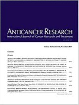
"Characterization of Pancreatic and Biliary Cancer Stem Cells in Patient-derived Tissue" was published in Anticancer Research. Authors are J. Gogolok, E. Seidel, A. Strönisch, A. Reutzel-Selke, I.M. Sauer, J. Pratschke, M. Bahra, and R.B. Schmuck.

