News

M. Pflüger: Resect or not to resect: Unravelling the Biology of Neoplastic Pancreatic Cysts Using Genetic Studies
Stacks Image 23717

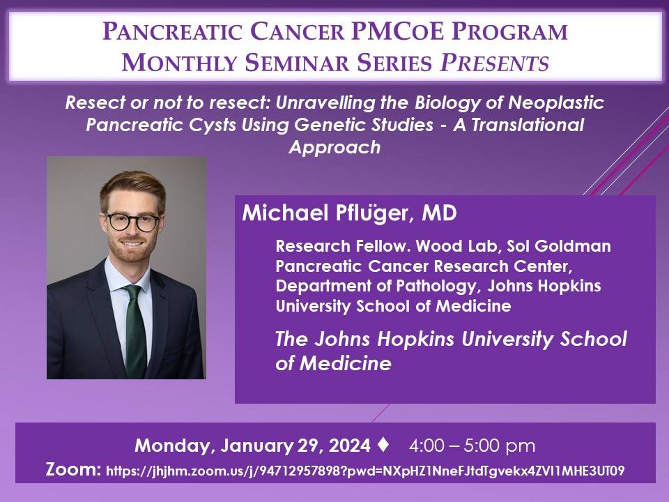
Michael Pflüger - currently research fellow at the Wood Lab, Sol Goldman Pancreatic Cancer Research Center, Department of Pathology, Johns Hopkins University School of Medicine - will present the translational work of the Wood Laboratory and the interdisciplinary management of pancreatic neoplasia at Johns Hopkins Hospital as part of the monthly interdisciplinary event series "The Pancreatic Cancer Precision Medicine Center of Excellence Program (PMCoE) Seminar Series".

"Resect or not to resect: Unravelling the Biology of Neoplastic Pancreatic Cysts Using Genetic Studies - A Translational Approach"
29.01.2024, 22:00/10 pm CET
Zoom link: https://jhjhm.zoom.us/j/94712957898?pwd=NXpHZ1NneFJtdTgvekx4ZVI1MHE3UT09
New DFG research group FOR 5628 with our participation
Stacks Image 23768
The Deutsche Forschungsgemeinschaft (DFG, German Research Foundation) is establishing eight new research groups. One of these groups is FOR 5628: "Multiscale magnetic resonance elastography in cancer: The mechanical niche of tumor formation and metastatic spread – towards an improved diagnosis of cancer through mechanical imaging". The speaker and initiator is Prof. Ingolf Sack.

During the development of a tumour, the tissue changes its shape, e.g., alternating between hard and fluidic states. For this, cells exert forces and are simultaneously influenced by forces. This research group is investigating which mechanical-physical processes are behind this. How do tumours and metastases develop? What makes them resistant to therapy? The team is investigating these questions using magnetic resonance elastography (MRE) – a new clinical procedure that can be used to record the mechanical properties of body tissue. The goal is to be able to better diagnose tumours.
Dr. Karl Hillebrandt and Prof. Dr. Igor Sauer are part of the research group as PI in three projects:
  • A03 Cancer cell unjamming and jamming as prerequisites for the formation of primary and metastatic tumors
  • B03 Scaffold composition and fluid pressure in recellularized hepatic and pancreatic tumors
  • C01 Multiscale mechanical properties of tumors and tumor environment – from tissue specimens to patients

Was are happy to be part of this exzellent team!
Work with us | PhD position
We offer a funded PhD position for a computational biologist @ Experimental Surgery, Charité – Universitätsmedizin Berlin!

Are you interested to work on cutting-edge cancer research, and investigating complex-chromosomal as well as other genomic phenomena in human carcinoma cells ?

  • You will work on complex genome analyses (single-cell analyses, whole genome analyses) to detect, annotate and re-construct circular DNA using state of the art computational tools (e.g., Amplicon architect).
  • We provide motivation and high commitment in supervision in areas around experimental oncology and surgery.
  • The position allows the applicant to pursue an academic qualification, while collaborating and networking with international experts on extrachromosomal circular DNA and pancreatic carcinogenesis.

Apply now!
Send brief cover letter and CV via email to Dr. med. Matthäus Felsenstein (matthaeus.felsenstein@charite.de)
BIH Medical Student Research Stipend for Cao Zhong Jing Jin
Stacks Image 23855
Cao Zhong Jing Jin under the supervision of Dr. med. Matthäus Felsenstein successfully applied for the BIH Medical Student Research Stipend on their project “Deciphering the molecular determinants for the transformation of high-grade pancreatic duct dysplasia to invasive carcinoma by single-cell transcriptomics”. She is conducting a cutting-edge research project merging molecular data with histological information in collaboration with the BIH core facilities for single-cell genomics (Dr. Thomas Conrad) and intelligent imaging (Prof. Dr. Christian Conrad). Using modern single-cell- and spatial transcriptomics on pancreatic precursor and carcinoma samples, the project aims at defining molecular signatures that drive dysplastic cells.

Congratulations!
EKFS grant for functional role and clinical relevance of ecDNA in pancreatic adenocarcinoma
Stacks Image 23875
Dr. Matthäus Felsenstein successfully applied for funding from the Else Kröner Fresenius Stiftung (EKFS) for his project “The functional role and clinical relevance of extrachromosomal DNA (ecDNA) in pancreatic adenocarcinoma”. In close collaboration with the excellence group around Professor Anton Henssen, he is aiming at improved understanding of unique genomic patterns as a results of complex chromosomal rearrangements in pancreatic adenocarcinoma that could drive its aggressive behavior. He will use state-of-the art three-dimensional tissue culture to enrich for neoplastic cells from primary PDAC specimen and subsequently perform genome analyses to identify samples that harbor extrachromosomal DNA. The clinical impact of these chromosomal structures will be explored by clinical correlation analyses and therapy response in vitro.

Congratulations!
Solid fraction determines stiffness and viscosity in decellularized pancreatic tissues
Stacks Image 23962
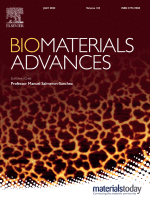
The article „Solid fraction determines stiffness and viscosity in decellularized pancreatic tissues“ in Biomaterials Advances is now available online.
There is free access to a PDF of the article here until August 20, 2022!

The role of extracellular matrix (ECM) composition and turnover in mechano-signaling and the metamorphic fate of cells seeded into decellularized tissue can be elucidated by recent developments in non-invasive imaging and biotechnological analysis methods. Because these methods allow accurate quantification of the composition and structural integrity of the ECM, they can be critical in establishing standardized decellularization protocols. This study proposes quantification of the solid fraction, the single-component fraction and the viscoelasticity of decellularized pancreatic tissues using compact multifrequency magnetic resonance elastography (MRE) to assess the efficiency and quality of decellularization protocols. MRE of native and decellularized pancreatic tissues showed that viscoelasticity parameters depend according to a power law on the solid fraction of the decellularized matrix. The parameters can thus be used as highly sensitive markers of the mechanical integrity of soft tissues. Compact MRE allows consistent and noninvasive quantification of the viscoelastic properties of decellularized tissue. Such a method is urgently needed for the standardized monitoring of decellularization processes, evaluation of mechanical ECM properties, and quantification of the integrity of solid structural elements remaining in the decellularized tissue matrix.

Authors are Joachim Snellings, Eriselda Keshi, Peter Tang, Assal Daneshgar, Esther C. Willma, Luna Haderer, Oliver Klein, Felix Krenzien, Thomas Malink, Patrick Asbach, Johann Pratschke, Igor M. Sauer, Jürgen Braun, Ingolf Sack, and Karl Hillebrandt.

Inaugural Lectures
We are pleased to announce that four members of staff have successfully completed their habilitation work in the last few months!

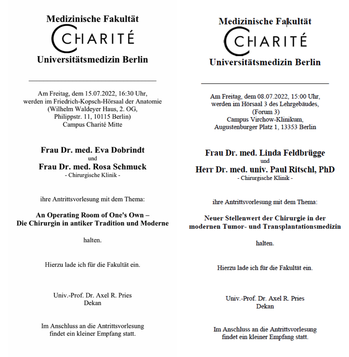
On
Friday, 08.07.2022 at 15:00 in lecture hall 3 of the teaching building (Forum 3, CVK), Dr. med. habil. Linda Feldbrügge and Dr. med. habil. Paul Ritschl will give their inaugural lectures entitled "New role of surgery in modern tumour and transplant medicine".

On
Friday, 15.07.2022 at 16:30 in the Friedrich Kopsch lecture theatre of the Anatomy Department at Campus Mitte Dr. med. habil. Eva Dobrindt and Dr. med. habil. Rosa Schmuck will present their inaugural lectures with the topic "An Operating Room of One's Own - The Surgeon in Ancient Tradition and Modernity".
This will be followed by a small reception in the park in front of the venue.
Stacks Image 23972
Two new research grants by Berliner Krebsgesellschaft
Stacks Image 24043
The Berliner Krebsgesellschaft will fund two very interesting research projects by Dr. Linda Feldbrügge and Dr. Karl Hillebrandt in collaboration with Dr. Björn Papke.

Stacks Image 24050
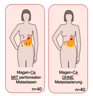
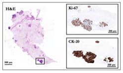
„Purinergic immune regulation in peritoneal metastases of gastric cancer via CD39 and ENTPD3 – target for a novel immune Checkpoint inhibition?“ – PI: Dr. Linda Feldbrügge

Peritoneal metastasis, especially derived from gastric cancer (GC), has a poor prognosis with a median survival of only months. Treatment is usually confined to palliative systemic chemotherapy, complemented individually by checkpoint inhibitors that block PD1-signaling. Innovative therapies combining surgery with local drug application such as hyperthermic intraperitoneal chemotherapy (HIPEC) or pressurized intraperitoneal aerosol chemotherapy (PIPAC) are still pending confirmation in clinical trials. Purinergic signaling, which involves ATP hydrolysis and generation of adenosine, regulated through CD39 (ENTPD1) and related enzymes, has been recognized as a critical immunoregulatory pathway in the tumor microenvironment (TME). The objective of the current project is to characterize the immune environment in the unique setting of peritoneal metastasis of gastric cancer with a focus on ectonucleotidases CD39 and ENTPD3 on T cells, macrophages and MDSC as well as mechanisms of ectonucleotidase-mediated immune regulation in tumor associated macrophages in vitro. As a high-volume center for surgical therapy of peritoneal malignancies and with years of experience in ectonucleotidase research, we aim to advance the understanding of peritoneal metastasis and contribute to improving treatment options for our patients.

Stacks Image 24057
"The influence of decellularised tumour matrix heterogeneity in relation to KRAS/MAPK inhibition of in vitro colorectal liver metastases." PI: Dr. Karl Hillebrandt and Dr. Björn Papke (Dept. of Pathology)

Colorectal cancer (CRC) is the second leading cause of cancer-related death worldwide, with approximately 900,000 annual deaths. 30-50% of patients develop colorectal liver metastases (CRLM) during their disease. More than 50% of these tumors have mutations in the KRAS oncogene, making them usually poorly treatable. Despite multimodal therapy concepts have improved the outcome of these patients, a large proportion of patients suffer a recurrence of their disease. For better therapeutic concepts, we need to better understand the tumor biology and metastatic mechanisms of these diseases. In vitro models, such as two-dimensional cell culture, are primarily used for this purpose. These models can only reflect the physiological complexity to a limited extent. Recently, it was shown that the use of organ-specific and tumor-specific extracellular matrix (ECM) has an impact on the behavior of human CRC cell lines. Culture of cell lines with decellularized matrix resulted in cells adopting a metastatic cell state and forming significantly more metastases in a mouse model than cells cultured on plastic or collagen. The goal of our project is to study the growth (with and without inhibition of the RAS/MAPK signaling pathway) of patient-derived tumor organoids growing on different decellularized metastatic matrices (dMM) and decellularized liver matrices (dLM). These studies of tumor matrix heterogeneity are essential to define which starting materials, for in vitro modeling of our three-dimensional tumor organoid culture, can be used to develop the most physiological, personalized dLM/dMM-based CRLM in vitro model possible. Based on these results, we plan to conduct small-scale therapy evaluations for personalized tumor therapy using our in vitro dLM/dMM-based CRLM in the near future.

Congratulations!
BIH Charité Clinician Scientist Symposium in Honor and Memory of Duška Dragun
Stacks Image 24080
28 May 2021 - 29 May 2021
BIH Charité Clinician Scientist Symposium in Honor and Memory of Duška Dragun

The symposium is composed of several components: First and foremost, it will commemorate Prof. Duška Dragun, the former Director of the BIH Biomedical Innovation Academy (BIA) and Director of the BIH Charité Clinician Scientist Program, who passed away in December 2020, and will be joined by stakeholders from academia and science policy. In addition, there will be scientific sessions, which will form tandems of program fellows and invited speaker. During a digital certificate ceremony on the evening of 28 May 2021, some 50 alumni will be bid farewell. The event language is English.

When
28 and 29 May 2021
10:00 - 6:30 pm

How
Online Event (semi-digital)

Registration
To receive the login link please register here.
Advanced Clinician Scientists
Stacks Image 24085
Priv.-Doz. Dr. Nathanael Raschzok and Priv.-Doz. Dr. Felix Krenzien successfully applied for the BIH Charité Advanced Clinician Scientist Pilot Programme (AdCSP) in a highly competitive process.

The BIH Charité AdCSP is designed as a career-phase-specific, sustainable funding programme that aims to closely interlink individual and institutional funding. The primary goal of the programme is to simultaneously incentivise the fellows and recognise the permissive academic culture of the respective clinics or institutes. Like the BIH Charité Clinician Scientist Programme (CSP) and the "Digital Clinician Scientist Programme" (DCSP), which has been additionally funded by the DFG since 2019, it is intended to be open to all clinical disciplines and to offer multiple networking opportunities for the funded fellows and participating clinics and institutes.

Congratulations!
Grant provided by the Berliner Krebsgesellschaft e.V.
Stacks Image 24090
Dr. med. M. Felsenstein receives a grant provided by the Berliner Krebsgesellschaft e.V. for his project "Deciphering the molecular determinants of pancreatic duct dysplasia by analysis of single-cell transcriptomics (RNAseq) in precursor lesions".

Besides great advances in the molecular and genetic understanding of pancreatic duct adenocarcinoma (PDAC), this tumor entity remains particularly aggressive with dismal prognosis. Recent single-cell sequencing studies underline the eminent urgency to understand tumor heterogeneity in the setting of PDAC. More detailed knowledge about the molecular mechanisms of pancreatic cancer evolution, carcinogenesis and heterogeneity could direct ideas for earlier detection and more effective targeted therapies, also preventing disease recurrence. Future therapeutic approaches in precision medicine will likely focus on the disease relevant sub-populations, specifically driving cancer progression, dissemination and exerting tumor escape mechanisms. In-depth transcriptomic data of single carcinoma environmental cells and respective cell clusters may help to discover novel biomarkers, which can be clinically instrumented for earlier detection and putatively increase the fraction of patients, amenable to curatively intended therapies. This study aims to analyze sorted single cells of macro-dissected precursor and cancerous lesions of the pancreas by single nuclei RNA sequencing (snRNAseq). In this feasibility study, we will include 10 patients, who will undergo resection of the pancreas due to “worrisome” or malignant lesions. We will perform in-depth transcriptomic analysis of pancreatic dysplasia in order to broaden our understanding of the molecular mechanisms of pancreatic carcinogenesis.

Congratulations!
Notch Signaling Pathway in Pancreatobiliary Tumors
Stacks Image 24110
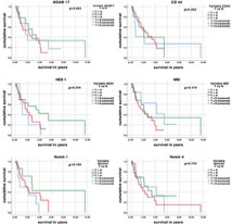
The Notch signaling pathway plays an important role both in the development of the ductal systems of the pancreas and the bile ducts as well as in cancer development and progression. The aim of this study was to examine the expression of central proteins of the Notch signaling pathway in pancreatobiliary tumors and its influence on patient survival.

Materials and Methods: We compared the receptors (Notch1, Notch4), activating splicing factors (ADAM17), and target genes (HES1) of the Notch pathway and progenitor cell markers with relevance for the Notch signaling pathway (CD44, MSI1) between pancreatic adenocarcinomas (PDAC, n = 14), intrahepatic cholangiocarcinoma (iCC, n = 24), and extrahepatic cholangiocarcinoma (eCC, n = 22) cholangiocarcinomas.

A significant overexpression of almost all studied components of the Notch signaling pathway can be found in the tumor tissue, however, without a significant influence on patient survival. Therefore, further studies are warranted to draw conclusions on Notch pathway's relevance for patient survival.

The paper "Notch Signaling Pathway in Pancreatobiliary Tumors" is available via Medicina, 2021;57(2):105. Authors are Francesca Borlak, Anja Reutzel-Selke, Anja Schirmeier, Julia Gogolok, Ellen von Hoerschelmann, Igor M Sauer, Johann Pratschke, Marcus Bahra, and Rosa B Schmuck.
Karl Hillebrandt | Charité 3R Tandem project for early career researchers
Stacks Image 24125
Together with Dr. Björn Papke (Molecular tumour pathology), Dr. Karl Hillebrandt was able to acquire funding for a "Tandem project for early career researchers" from the Charité 3R. The project is entitled "A personalised therapy approach implementing individually matched matrix-based in vitro colorectal liver metastases to reduce metastatic mouse models".
Although modern multimodal therapy strategies have improved the clinical outcome of patients with colorectal liver metastases (CRLM), the overall prognosis is still poor. To further improve treatment options for patients, it is necessary to develop and test new targeted therapeutic approaches. To date, mouse models have often been used to study metastatic colorectal cancer. However, the rate of successful translation of animal models into clinical trials is less than 8%, highlighting the urgent need for alternative models to study the biology of metastatic cancer. This project aims to develop a novel personalised extracellular matrix-based in vitro model of human CRLM. This model will be validated against existing data from patient-derived organoids and xenografts (histology, single cell RNA sequencing and targeted gene sequencing). After internal comparison of our in vitro CRLM with the original CRLM, we will translate it into a personalised drug screening platform to test drug response from standard therapy to novel inhibitor combinations.
Characterization of pancreatic and biliary cancer stem cells in patient-derived tissue
Stacks Image 24247
Pancreatic ductal adenocarcinoma (PDAC) and extrahepatic cholangio-carcinoma (eCC) represent two cancer entities with devastating prognoses. Despite recent progress in research and treatment, therapy remains challenging. Cancer stem cells (CSCs) have been shown to play an important role in metastasis and chemoresistance. Therefore, CSCs may play a promising role as a potential therapeutic target.
A total of 31 patients (23 PDAC, 8 eCC) were included in the study. CSCs were analyzed in a single-cell suspension of tumor samples via fluorescence-activated cell scanning (FACS) with a functional Hoechst 33342 staining as well as a cell surface marker staining of the CSC-panel (CD24, CD44 and EpCAM) and markers to identify fibroblasts, leukocytes and components of the notch signaling pathway. Furthermore, the potential presence of CSCs among primary cancer-associated fibroblasts (CAFs) was assessed using the same FACS-panel.
We showed that CSCs are present in patient-derived dissociated tumor tissue. The functional and surface marker profile of CSC-detection did in fact correlate. The amount of CSCs was significantly correlated with tumor characteristics such as a higher UICC stadium and nodal invasion. CSCs were not restricted to the epithelial cell fraction in tumor tissues, which has been verified in independent analysis of primary cell cultures of CAFs.
Our study confirms the in vivo presence of CSCs in PDAC and eCC, stating a clinical significance thereof and thus their plausibility as therapeutic targets. In addition, stem-like cells also seem to constitute a part of the CAFs.

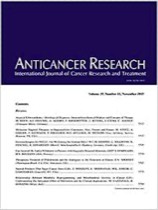
"Characterization of Pancreatic and Biliary Cancer Stem Cells in Patient-derived Tissue" was published in Anticancer Research. Authors are J. Gogolok, E. Seidel, A. Strönisch, A. Reutzel-Selke, I.M. Sauer, J. Pratschke, M. Bahra, and R.B. Schmuck.
Two new BIH Charité Junior Clinician Scientists
Stacks Image 24309
Dr. Karl Hillebrandt and Dr. Matthäus Felsenstein successfully applied for the BIH Charité Junior Clinician Scientist Program. Karl Hillebrandt will continue his work on human decellularized liver slices as 3D platform for in vitro models of cholangiocellular carcinoma. Matthäus Felsenstein focusses on derivation of normal pancreatic duct cells from human primary tissue and their stepwise genetic modification in vitro using CRISPR/Cas9 .

Congratulations!
 Page 1 / 1 
M. Pflüger: Resect or not to resect: Unravelling the Biology of Neoplastic Pancreatic Cysts Using Genetic Studies
Stacks Image 19713

Michael Pflüger - currently research fellow at the Wood Lab, Sol Goldman Pancreatic Cancer Research Center, Department of Pathology, Johns Hopkins University School of Medicine - will present the translational work of the Wood Laboratory and the interdisciplinary management of pancreatic neoplasia at Johns Hopkins Hospital as part of the monthly interdisciplinary event series "The Pancreatic Cancer Precision Medicine Center of Excellence Program (PMCoE) Seminar Series".

"Resect or not to resect: Unravelling the Biology of Neoplastic Pancreatic Cysts Using Genetic Studies - A Translational Approach"
29.01.2024, 22:00/10 pm CET
Zoom link: https://jhjhm.zoom.us/j/94712957898?pwd=NXpHZ1NneFJtdTgvekx4ZVI1MHE3UT09
Stacks Image 27655
Our manuscript "Depletion of donor dendritic cells ameliorates immunogenicity of both skin and hind limb transplants" has been accepted for publication in Frontiers in Immunology, section Alloimmunity and Transplantation. Authors are Muhammad Imtiaz Ashraf, Joerg Mengwasser, Anja Reutzel-Selke, Dietrich Polenz, Kirsten Führer, Steffen Lippert, Peter Tang, Edward Michaelis, Rusan Catar, Johann Pratschke, Christian Witzel, Igor M. Sauer, Stefan G. Tullius, and Barbara Kern.

Acute cellular rejection remains a significant obstacle affecting successful outcomes of organ transplantation including vascularized composite tissue allografts (VCA). Donor antigen presenting cells (APC), particularly dendritic cells (DC), orchestrate early alloimmune responses by activating recipient effector T cells. Employing a targeted approach, we investigated the impact of donor-derived conventional DC (cDC) and APC on the immunogenicity of skin and skin-containing VCA grafts, using mouse models of skin and hind limb transplantation.
By post-transplantation day 6, skin grafts demonstrated severe rejections, characterized by predominance of recipient CD4 T cells. In contrast, hind limb grafts showed moderate rejection, primarily infiltrated by CD8 T cells. While donor depletion of cDC and APC reduced frequencies, maturation, and activation of DC in all analysed tissues of skin transplant recipients, reduction in DC activities was only observed in the spleen of hind limb recipients. Donor cDC and APC depletion did not impact all lymphocyte compartments but significantly affected CD8 T cells and activated CD4 T in lymph nodes of skin recipients. Moreover, both donor APC and cDC depletion attenuated the Th17 immune response, evident by significantly reduced Th17 (CD4+IL-17+) cells in the spleen of skin recipients and reduced levels of IL-17E and lymphotoxin-α in the serum samples of both skin and hind limb recipients. In conclusion, our findings underscore the highly immunogenic nature of skin component in VCA. The depletion of donor APC and cDC mitigates the immunogenicity of skin grafts while exerting minimal impact on VCA.

Archive


Categories

Year

This website or its third-party tools use cookies, which are necessary to its functioning and required to achieve the purpose illustrated in the Disclaimer. By closing this banner, scrolling this page, clicking a link or continuing to browse otherwise, you agree to the use of cookies.