News

Proteomic analysis of decellularized mice liver and kidney extracellular matrices
Stacks Image 23711
Based on the collaboration between the Department of General, Visceral, and Transplant Surgery, University Hospital Münster, and Experimental Surgery, Department of Surgery, Charité – Universitätsmedizin Berlin our work on the "Proteomic analysis of decellularized mice liver and kidney extracellular matrices" has been accepted for publication in Journal of Biological Engineering.

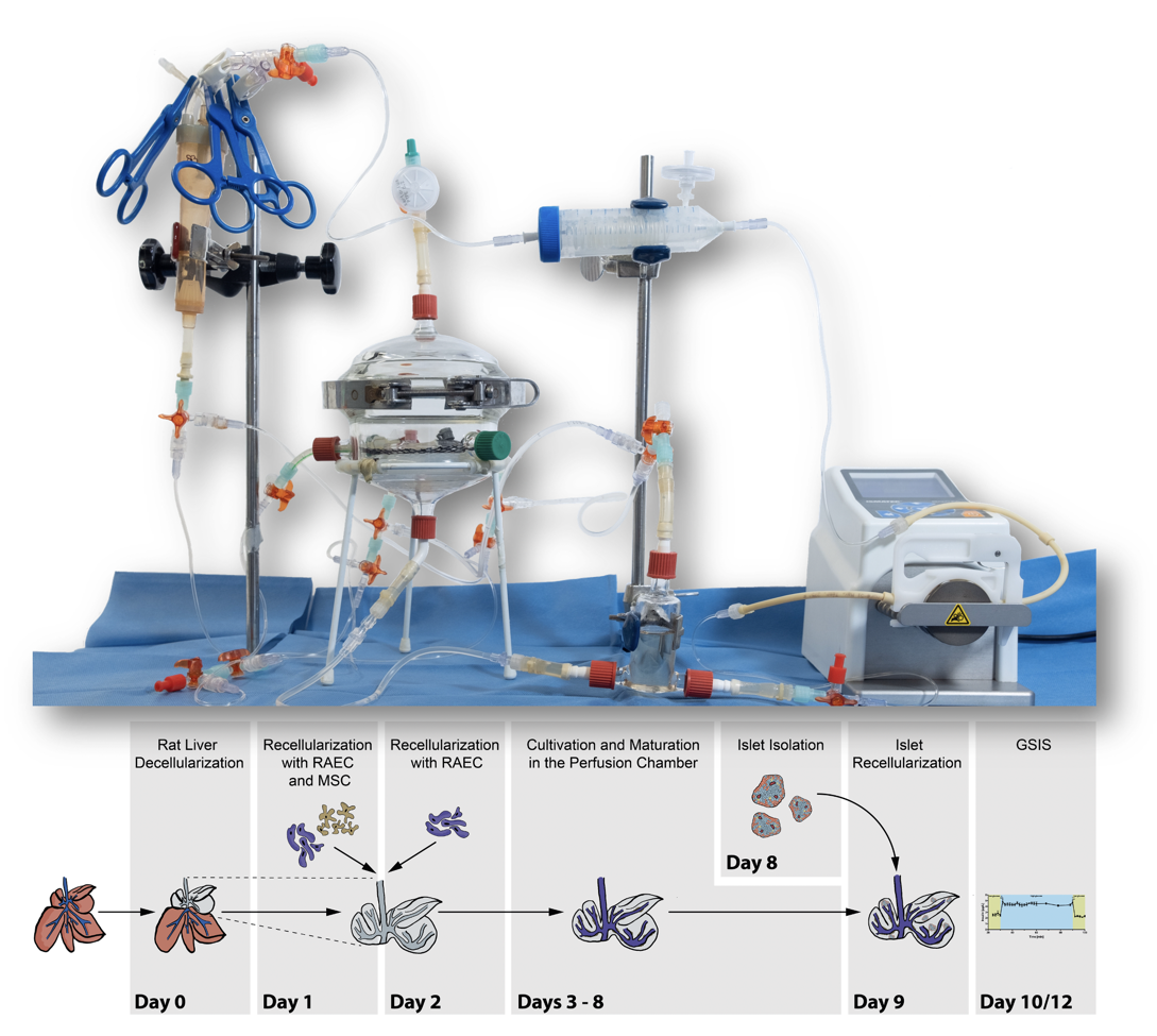
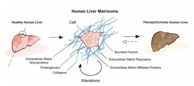
In this study, we employed a bottom-up proteomic approach to elucidate the intricate network of proteins in the decellularized extracellular matrices of murine liver and kidney tissues. This approach involved the use of a novel, perfusion-based decellularization protocol to generate acellular whole organ scaffolds. Proteomic analysis of decellularized mice liver and kidney ECM scaffolds revealed tissue-specific differences in matrisome composition, while we found a predominantly stable composition of the core matrisome, consisting of collagens, glycoproteins, and proteoglycans. Liver matrisome analysis revealed unique proteins such as collagen type VI alpha-6, fibrillin-2 or biglycan. In the kidney, specific ECM-regulators such as cathepsin z were detected. The identification of distinct proteomic signatures provides insights into how different matrisome compositions might influence the biological properties of distinct tissues. This experimental workflow will help to further elucidate the proteomic landscape of decellularized extracellular matrix scaffolds of mice in order to decipher complex cell-matrix interactions and their contribution to a tissue-specific microenvironment.

Authors are Anna-Maria Diedrich, Assal Daneshgar, Peter Tang, Oliver Klein, Annika Mohr, Olachi A. Onwuegbuchulam, Sabine von Rueden, Kerstin Menck, Annalen Bleckmann, Mazen A. Juratli, Felix Becker, Igor M. Sauer, Karl H. Hillebrandt, Andreas Pascher, and Benjamin Struecker.
Engineering an endothelialized, endocrine NeoPancreas
Stacks Image 24164
Acta Biomaterialia accepted our latest paper on "Engineering an endothelialized, endocrine Neo-Pancreas: evaluation of islet functionality in an ex vivo model".

Islet-based recellularization of decellularized, repurposed rat livers may form a transplantable Neo-Pancreas. The aim of this study is the establishment of the necessary protocols, the evaluation of the organ structure and the analysis of the islet functionality ex vivo.
After perfusion-based decellularization of rat livers, matrices were repopulated with endothelial cells and mesenchymal stromal cells, incubated for 8 days in a perfusion chamber and finally repopulated on day 9 with intact rodent islets. Integrity and quality of re-endothelialization was assessed by histology and FITC-dextran perfusion assay. Functionality of the islets of Langerhans was determined on day 10 and day 12 via glucose stimulated insulin secretion.
Blood gas analysis variables confirmed the stability of the perfusion cultivation. Histological staining showed that cells formed a monolayer inside the intact vascular structure. These findings were confirmed by electron microscopy. Islets infused via the bile duct could histologically be found in the parenchymal space. Adequate insulin secretion after glucose stimulation after 1-day and 3-day cultivation verified islet viability and functionality after the repopulation process.
We provide the first proof-of-concept for the functionality of islets of Langerhans engrafted in a decellularized rat liver. Furthermore, a re-endothelialization step was implemented to provide implantability. This technique can serve as a bioengineered platform to generate implantable and functional endocrine Neo-Pancreases.

Authors are Hannah Everwien, Eriselda Keshi, Karl H. Hillebrandt, Barbara Ludwig, Marie Weinhart, Peter Tang, Anika S. Beierle, Hendrik Napierala, Joseph MGV Gassner, Nicolai Seiffert, Simon Moosburner, Dominik Geisel, Anja Reutzel-Selke, Benjamin Strücker, Johann Pratschke, Nils Haep, and Igor M. Sauer.
Stacks Image 24166
The Human Liver Matrisome
Stacks Image 24191
Biomaterials accepted our latest paper on „The Human Liver Matrisome – Proteomic Analysis of Native and Fibrotic Human Liver Extracellular Matrices for Organ Engineering Approaches“.

The production of biomaterials that endow significant morphogenic and microenvironmental cues for the constitution of cell integration and regeneration remains a key challenge in the successful implementation of functional organ replacements. Despite the vast development in the production of biological and architecturally native matrices, the complex compositions and pivotal figures by which the human matrisome mediates many of its essential functions are yet to be defined. Here we present a thorough analysis of the native human liver proteomic landscape using decellularization and defatting protocols to extract create extracellular matrix scaffolds of natural origin that can further be used in both bottom-up and top-down approaches in tissue engineering based organ replacements. Furthermore, by analyzing human liver extracellular matrices in different stages of fibrosis and cirrhosis, we have identified distinct attributes of these tissues that could potentially be exploited therapeutically and thus require further investigation. The general experimental pipeline presented in this study is applicable to any type of tissue and can be widely used for different approaches in regenerative medicine and in the construction of novel biomaterials for organ engineering approaches.

Authors are A. Daneshgar, O. Klein, G. Nebrich, M. Weinhart, P. Tang, A. Arnold, I. Ullah, J. Pohl, S. Moosburner, N. Raschzok, B. Strücker, M. Bahra, J. Pratschke, I.M. Sauer, and K.H. Hillebrandt. The authors acknowledge the support of the Cluster of Excellence Matters of Activity. Image Space Material funded by the Deutsche Forschungsgemeinschaft (DFG, German Research Foundation) under Germany´s Excellence Strategy – EXC 2025.
Stacks Image 24193
Magnetic resonance elastography quantification of decellularized liver tissue
Stacks Image 24242
"Magnetic resonance elastography quantification of the solid-to-fluid transition of liver tissue due to decellularization" was published in the latest issue of the Journal of the Mechanical Behavior of Biomedical Materials.

Maintenance of tissue extracellular matrix (ECM) and its biomechanical properties for tissue engineering is one of the substantial challenges in the field of decellularization and recellularization. Preservation of the organ-specific biomatrix is crucial for successful recellularization to support cell survival, proliferation, and functionality. However, understanding ECM properties with and without its inhabiting cells as well as the transition between the two states lacks appropriate test methods capable of quantifying bulk viscoelastic parameters in soft tissues.
We used compact magnetic resonance elastography (MRE) with 400, 500, and 600 Hz driving frequency to investigate rat liver specimens for quantification of viscoelastic property changes resulting from decellularization. Tissue structures in native and decellularized livers were characterized by collagen and elastin quantification, histological analysis, and scanning electron microscopy.
Decellularization did not affect the integrity of microanatomy and structural composition of liver ECM but was found to be associated with increases in the relative amounts of collagen by 83-fold (37.4 ± 17.5 vs. 0.5 ± 0.01 μg/mg, p = 0.0002) and elastin by approx. 3-fold (404.1 ± 139.6 vs. 151.0 ± 132.3 μg/mg, p = 0.0046). Decellularization reduced storage modulus by approx. 9-fold (from 4.9 ± 0.8 kPa to 0.5 ± 0.5 kPa, p < 0.0001) and loss modulus by approx. 7-fold (3.6 kPa to 0.5 kPa, p < 0.0001), indicating a marked loss of global tissue rigidity as well as a property shift from solid towards more fluid tissue behavior (p = 0.0097).
Our results suggest that the rigidity of liver tissue is largely determined by cellular components, which are replaced by fluid-filled spaces when cells are removed. This leads to an overall increase in tissue fluidity and a viscous drag within the relatively sparse remaining ECM. Compact MRE is an excellent tool for quantifying the mechanical properties of decellularized biological tissue and a promising candidate for useful applications in tissue engineering.

Authors are Hannah Everwien, Angela Ariza de Schellenberger, Nils Haep, Heiko Tzschätzsch, Johann Pratschke, Igor M. Sauer, Jürgen Braun, Karl H. Hillebrandt and Ingolf Sack.

J Mech Behav Biomed Mater. 2020 Apr;104:103640. doi: 10.1016/j.jmbbm.2020.103640. Epub 2020 Jan 14.
New book: Decellularized Extracellular Matrix: Characterization, Fabrication and Applications
Stacks Image 24257
The extracellular matrix (ECM) supports cells and regulates various cellular functions in our body. The native ECM promises to provide an excellent scaffold for regenerative medicine. In order to use the ECM as a scaffold in medicine, its cellular fractions need to be removed while retaining its structural and compositional properties. This process is called decellularization, and the resulting product is known as the decellularized extracellular matrix (dECM).
The book Decellularized Extracellular Matrix: Characterization, Fabrication and Applications (Editors: Tetsuji Yamaoka, Takashi Hoshiba) focuses on the sources of dECM and its preparation, characterization techniques, fabrication, and applications in regenerative medicine and biological studies. Using this book, the reader will gain a good foundation in the field of ECM decellularization complemented with a broad overview of dECM characterization, ranging from structural observation and compositional assessment to immune responses against dECM and applications, ranging from microfabrication and 3D-printing to the application of tissue-derived dECM in vascular grafts and corneal tissue engineering etc. The book closes with a section dedicated to cultured cell dECM, a complementary technique of tissue-derived dECM preparation, for application in tissue engineering and regenerative medicine, addressing its use in stem cell differentiation and how it can help in the study of the tumor microenvironment as well as in clinical trials of peripheral nerve regeneration.

E. Keshi, I.M. Sauer and K.H. Hillebrandt contributed the chapter "Engineering an endocrine Neo-Pancreas".

The print version of this book (Royal Society of Chemistry, ISBN 978-1-78801-467-0) is planned for release on 11 December 2019.
 Page 1 / 1 
Stacks Image 27655
Our manuscript "Depletion of donor dendritic cells ameliorates immunogenicity of both skin and hind limb transplants" has been accepted for publication in Frontiers in Immunology, section Alloimmunity and Transplantation. Authors are Muhammad Imtiaz Ashraf, Joerg Mengwasser, Anja Reutzel-Selke, Dietrich Polenz, Kirsten Führer, Steffen Lippert, Peter Tang, Edward Michaelis, Rusan Catar, Johann Pratschke, Christian Witzel, Igor M. Sauer, Stefan G. Tullius, and Barbara Kern.

Acute cellular rejection remains a significant obstacle affecting successful outcomes of organ transplantation including vascularized composite tissue allografts (VCA). Donor antigen presenting cells (APC), particularly dendritic cells (DC), orchestrate early alloimmune responses by activating recipient effector T cells. Employing a targeted approach, we investigated the impact of donor-derived conventional DC (cDC) and APC on the immunogenicity of skin and skin-containing VCA grafts, using mouse models of skin and hind limb transplantation.
By post-transplantation day 6, skin grafts demonstrated severe rejections, characterized by predominance of recipient CD4 T cells. In contrast, hind limb grafts showed moderate rejection, primarily infiltrated by CD8 T cells. While donor depletion of cDC and APC reduced frequencies, maturation, and activation of DC in all analysed tissues of skin transplant recipients, reduction in DC activities was only observed in the spleen of hind limb recipients. Donor cDC and APC depletion did not impact all lymphocyte compartments but significantly affected CD8 T cells and activated CD4 T in lymph nodes of skin recipients. Moreover, both donor APC and cDC depletion attenuated the Th17 immune response, evident by significantly reduced Th17 (CD4+IL-17+) cells in the spleen of skin recipients and reduced levels of IL-17E and lymphotoxin-α in the serum samples of both skin and hind limb recipients. In conclusion, our findings underscore the highly immunogenic nature of skin component in VCA. The depletion of donor APC and cDC mitigates the immunogenicity of skin grafts while exerting minimal impact on VCA.

Archive


Categories

Year

This website or its third-party tools use cookies, which are necessary to its functioning and required to achieve the purpose illustrated in the Disclaimer. By closing this banner, scrolling this page, clicking a link or continuing to browse otherwise, you agree to the use of cookies.