Funding for "Expansion of Clinical Applications for the Human Muscle Stem Cell Product PHSat"
Stacks Image 28256
The Investitionsbank Berlin (IBB) is funding the collaborative project "Expansion of Clinical Applications for the Human Muscle Stem Cell Product PHSat", initiated by the start-up company MyoPax in cooperation with the Departments of Surgery and Neurosurgery at the Charité.
 
Muscle diseases affect more than 20 million patients in Europe with limited therapeutic options. MyoPax, a spin-off of Charité and MDC, has developed a patented method to isolate and expand patient-specific muscle stem cell populations, known as PHSats (Primary Human Satellite-cell derived muscle stem cells), while preserving their regenerative potential.
 
The ProFit project aims to preclinically evaluate additional clinical applications for PHSats. The validation of new indications will lay the foundation for future clinical studies and the expansion of the market potential of PHSats. In a subproject led by Experimental Surgery and PI Priv.-Doz. Dr. Karl Hillebrandt, the preclinical application of decellularized diaphragms combined with PHSats to regenerate the impaired diaphragms of mice with muscular dystrophy will be explored. Two routes of administration will be investigated: direct injection into damaged diaphragms and transplantation onto degenerated diaphragms. The ultimate goal is to preserve or improve respiratory function by augmenting the diaphragm. This approach holds promise for several conditions, such as ventilator-induced diaphragm dysfunction or diaphragm atrophy due to COPD, where impaired diaphragm function affects respiratory capacity.
Stacks Image 28258
Surgical planning in virtual reality: a systematic review
Stacks Image 28265
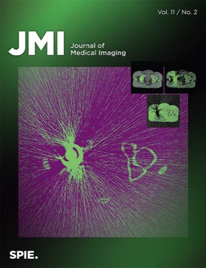
We just published a review on surgical planning in VR in the Journal of Medical Imaging. In the systematic review we look into how virtual reality (VR) is transforming surgical planning. With VR physicians can assess patient-specific image data in 3D, enhancing surgical decision-making and spatial localization of pathologies. We found that benefits of VR become more evident. However, its application in surgical planning remains experimental, with a need for refined study designs, improved technical reporting, and enhanced VR software usability for effective clinical implementation. Authors of "Surgical planning in virtual reality: a systematic review" are Prof. Dr. Moritz Queisner and Karl Eisenträger.

Virtual reality (VR) technology has emerged as a promising tool for physicians, offering the ability to assess anatomical data in 3D with visuospatial interaction qualities. This systematic review aims to provide an up-to-date overview of the latest research on VR in the field of surgical planning.
A comprehensive literature search was conducted based on the preferred reporting items for systematic reviews and meta-analyses covering the period from April 1, 2021 to May 10, 2023. The review summarizes the current state of research in this field, identifying key findings, technologies, study designs, methods, and potential directions for future research. Results show that the application of VR for surgical planning is still in an experimental stage but is gradually advancing toward clinical use. The diverse study designs, methodologies, and varying reporting hinder a comprehensive analysis. Some findings lack statistical evidence and rely on subjective assumptions. To strengthen evaluation, future research should focus on refining study designs, improving technical reporting, defining visual and technical proficiency requirements, and enhancing VR software usability and design. Addressing these areas could pave the way for an effective implementation of VR in clinical settings.
Spacial computing in the OR
Stacks Image 28271

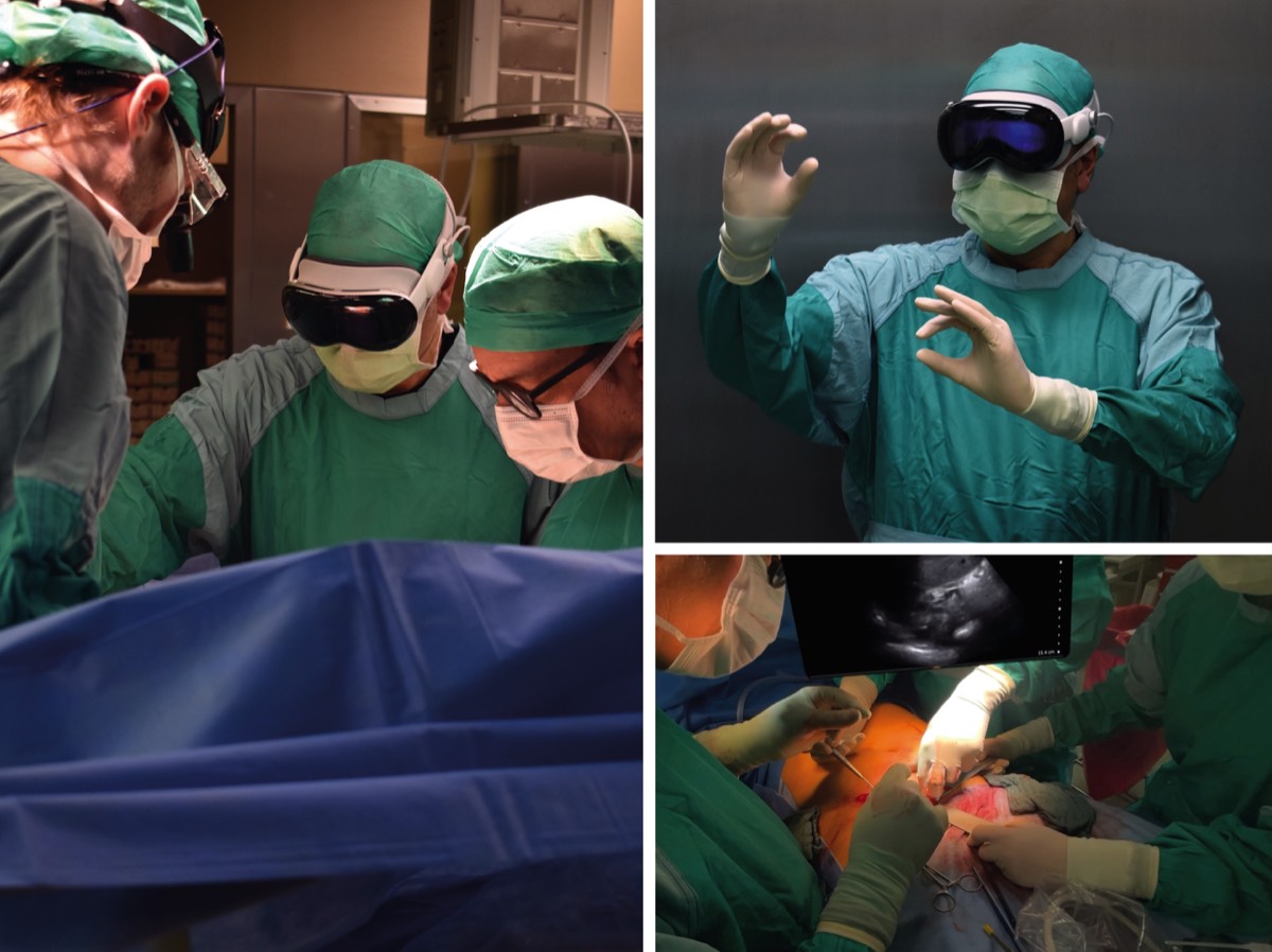
We tested the Apple Vision Pro in the operating theatre and it cuts an excellent figure: great images even in challenging lighting situations, stable interaction with the device - even though the limited peripheral vision and awareness inherent to video-based devices is a considerable downside in surgery.

We are looking forward to our first software solutions for improved hand-eye coordination in visceral surgery for this device too!
Optimizing environmental enrichment for Sprague Dawley rats: Exemplary insights into the liver proteome
Stacks Image 28280
Considering the expected increase in the elderly population and the growing emphasis on aging-related biomedical research, the demand for aged laboratory animals has surged, challenging established husbandry practices. Our objective was to establish a cost-effective method for environmental enrichment, utilizing the liver as a representative organ to assess metabolic changes in response to differing enrichment levels.
We conducted a six-month study involving 24 male Sprague Dawley rats who were randomly assigned to four environmental enrichment groups. Two groups were housed in standard cages, while the others were placed in modified rabbit cages. Half of the groups received weekly playtime in an enriched rat housing unit. We evaluated hormone levels, playtime behavior, and subjective handling experience. Additionally, liver tissue proteomic analysis was performed.
Initial corticosterone levels and those after 3 and 6 months showed no significant differences. Yet, testosterone levels were lower in the control group by the end of the study (p=0.007). In the liver tissue, we detected 1,871 distinct proteins, with 77% of them being consistent across all groups. In gene ontology analysis, no specific pathways were overexpressed. In semiquantitative analysis, we observed differences in proteins associated in lipid metabolism such as Apolipoprotein A-I and Acyl-CoA 6-desaturase, which were lower in the control group (p= 0.024 and p=0.009). Enriched environments reduced rat distress, large cages eased handling, and conflicts between rats lessened with bi-weekly interactions.

The manuscript "Optimizing environmental enrichment for Sprague Dawley rats: Exemplary insights into the liver proteome" has been accepted for publication in PLOS ONE.
Authors are Nathalie N. Roschke, Karl H. Hillebrandt, Dietrich Polenz, Oliver Klein, Joseph MGV Gassner, Johann Pratschke, Felix Krenzien, Igor M. Sauer, Nathanael Raschzok, and Simon Moosburner.
Proteomic analysis of decellularized mice liver and kidney extracellular matrices
Stacks Image 28285
Based on the collaboration between the Department of General, Visceral, and Transplant Surgery, University Hospital Münster, and Experimental Surgery, Department of Surgery, Charité – Universitätsmedizin Berlin our work on the "Proteomic analysis of decellularized mice liver and kidney extracellular matrices" has been accepted for publication in Journal of Biological Engineering.

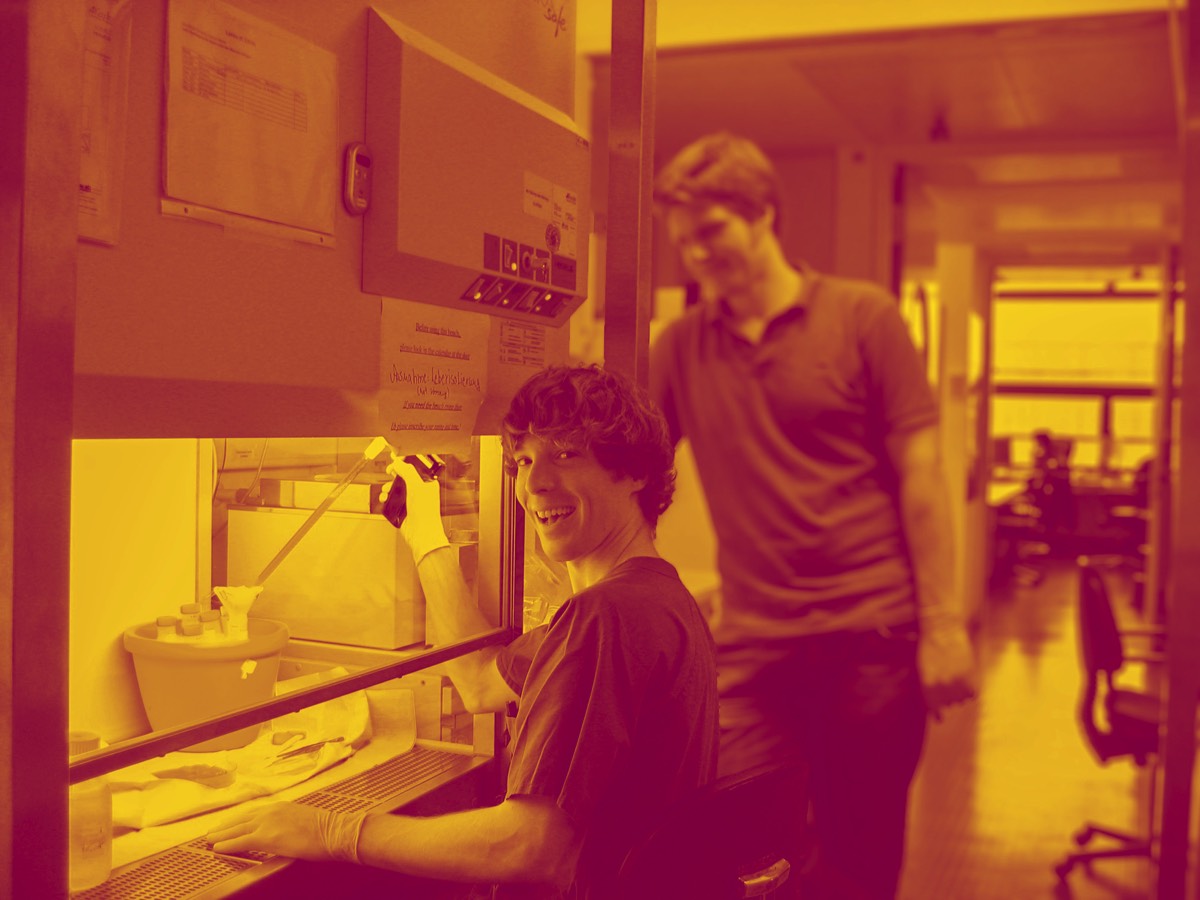
In this study, we employed a bottom-up proteomic approach to elucidate the intricate network of proteins in the decellularized extracellular matrices of murine liver and kidney tissues. This approach involved the use of a novel, perfusion-based decellularization protocol to generate acellular whole organ scaffolds. Proteomic analysis of decellularized mice liver and kidney ECM scaffolds revealed tissue-specific differences in matrisome composition, while we found a predominantly stable composition of the core matrisome, consisting of collagens, glycoproteins, and proteoglycans. Liver matrisome analysis revealed unique proteins such as collagen type VI alpha-6, fibrillin-2 or biglycan. In the kidney, specific ECM-regulators such as cathepsin z were detected. The identification of distinct proteomic signatures provides insights into how different matrisome compositions might influence the biological properties of distinct tissues. This experimental workflow will help to further elucidate the proteomic landscape of decellularized extracellular matrix scaffolds of mice in order to decipher complex cell-matrix interactions and their contribution to a tissue-specific microenvironment.

Authors are Anna-Maria Diedrich, Assal Daneshgar, Peter Tang, Oliver Klein, Annika Mohr, Olachi A. Onwuegbuchulam, Sabine von Rueden, Kerstin Menck, Annalen Bleckmann, Mazen A. Juratli, Felix Becker, Igor M. Sauer, Karl H. Hillebrandt, Andreas Pascher, and Benjamin Struecker.
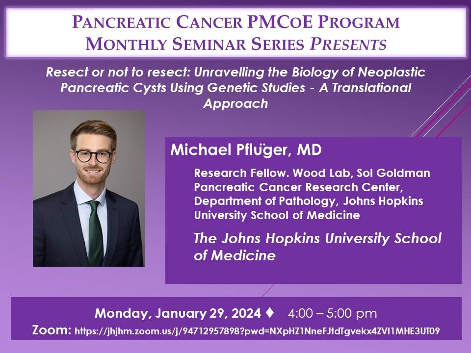
M. Pflüger: Resect or not to resect: Unravelling the Biology of Neoplastic Pancreatic Cysts Using Genetic Studies
Stacks Image 28291

Michael Pflüger - currently research fellow at the Wood Lab, Sol Goldman Pancreatic Cancer Research Center, Department of Pathology, Johns Hopkins University School of Medicine - will present the translational work of the Wood Laboratory and the interdisciplinary management of pancreatic neoplasia at Johns Hopkins Hospital as part of the monthly interdisciplinary event series "The Pancreatic Cancer Precision Medicine Center of Excellence Program (PMCoE) Seminar Series".

"Resect or not to resect: Unravelling the Biology of Neoplastic Pancreatic Cysts Using Genetic Studies - A Translational Approach"
29.01.2024, 22:00/10 pm CET
Zoom link: https://jhjhm.zoom.us/j/94712957898?pwd=NXpHZ1NneFJtdTgvekx4ZVI1MHE3UT09
miRNA as potential biomarkers after liver transplantation: A systematic review
Stacks Image 28304
The publication "miRNA as potential biomarkers after liver transplantation: A systematic review" is now available online in Transplantation Reviews. Authors are Pia F. Koch, Kristina Ludwig, Felix Krenzien, Karl H. Hillebrandt, Wenzel Schöning, Johann Pratschke, Nathanael Raschzok, Igor M. Sauer, and Simon Moosburner.
Early and accurate diagnosis of acute cellular rejection (ACR) and graft complications after liver transplantation is crucial for timely intervention and improved patient outcomes, but their diagnosis rely currently on invasive biopsy sampling, thus prompting the search for non-invasive Biomarkers. MicroRNA (miRNA) have emerged as promising biomarkers in various pathological conditions, and their potential utility in diagnosing acute cellular rejection after liver transplantation has gained significant interest.

This systematic review analyzes studies exploring miRNA as biomarkers for ACR and graft dysfunction in liver transplantation (PROSPERO ID CRD42023465278). The Cochrane Collaboration tool for assessing risk of bias was employed. Population data, identified miRNA and their dynamic regulation, as well as event prediction were compared. Data extraction and quality assessment were performed independently by two reviewers.

The results highlight the potential of miRNA as specific, non-invasive biomarkers for ACR and graft dysfunction following liver transplantation. However, further research is needed to validate these findings and establish standardized diagnostic panels to incorporate them into clinical practice and explore miRNA-based therapies in the future.
AI-based intra- and postoperative measurement from stereoimages
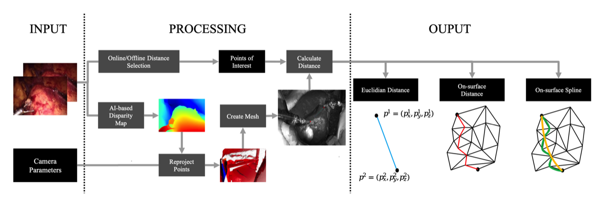
The publication "Redefining the Laparoscopic Spatial Sense: AI-based Intra- and Postoperative Measurement from Stereoimages“ has been accepted for the 38th AAAI Conference on Artificial Intelligence and is available via https://doi.org/10.48550/arXiv.2311.09744. The publication is the result of a fruitful collaboration between Karlsruhe Institute of Technology (KIT), Fraunhofer FIT, University of Bayreuth, and Charité – Universitätsmedizin Berlin. Authors are Leopold Müller, Patrick Hemmer, Moritz Queisner, Igor Sauer, Simeon Allmendinger, Johannes Jakubik, Michael Vössing, and Niklas Kühl.

A significant challenge in image-guided surgery is the accurate measurement task of relevant structures such as vessel segments, resection margins, or bowel lengths. While this task is an essential component of many surgeries, it involves substantial human effort and is prone to inaccuracies. In this paper, we develop a novel human-AI-based method for laparoscopic measurements utilizing stereo vision that has been guided by practicing surgeons. Based on a holistic qualitative requirements analysis, this work proposes a comprehensive measurement method, which comprises state-of-the-art machine learning architectures, such as RAFT-Stereo and YOLOv8. The developed method is assessed in various realistic experimental evaluation environments. Our results outline the potential of our method achieving high accuracies in distance measurements with errors below 1 mm. Furthermore, on-surface measurements demonstrate robustness when applied in challenging environments with textureless regions. Overall, by addressing the inherent challenges of image-guided surgery, we lay the foundation for a more robust and accurate solution for intra- and postoperative measurements, enabling more precise, safe, and efficient surgical procedures.

Stacks Image 28320
Priv.-Doz. Dr. med. Karl Hillebrandt
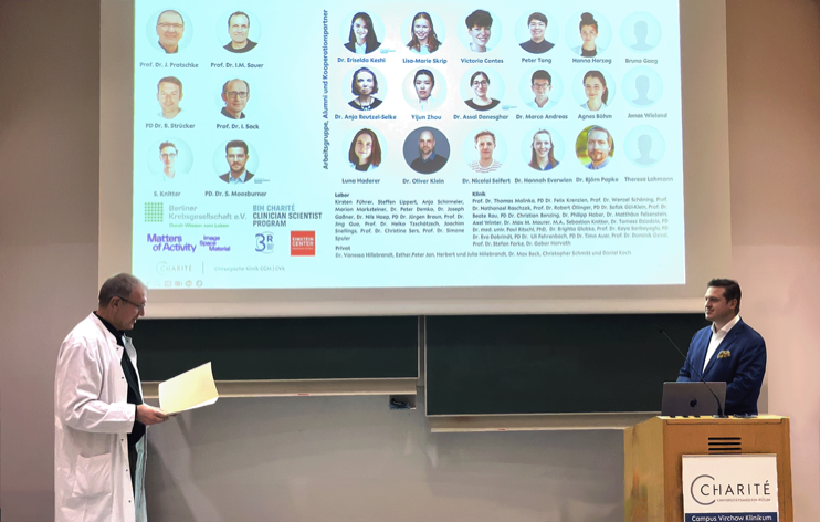
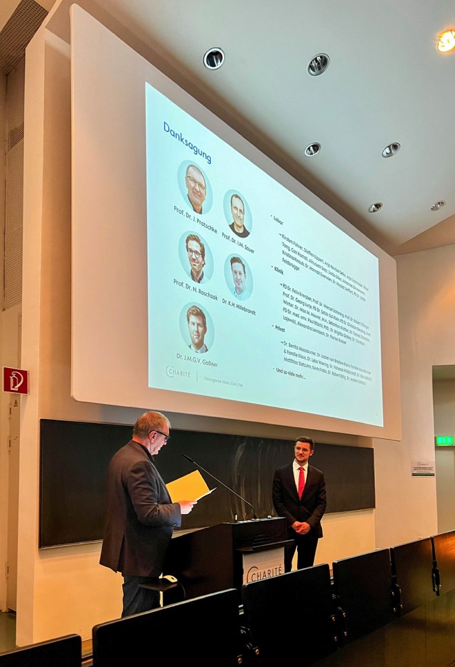
Today Karl Hillebrandt gave an exzellent inaugural lecture entitled „Die Geister, die ich rief ... – Eine kurze Geschichte der De- und Rezellularisierung“ and is now a private lecturer (Privatdozent) at the Charité – Universitätsmedizin Berlin and habilitated in the field of "Experimental Surgery".

He is being honored for his achievements in the field of tissue engineering. His postdoctoral thesis is entitled "New approaches for the characterisation of decellularised tissues and the recellularisation of vessels".

Congratulations!

Stacks Image 28330
Rise of tissue- and species-specific 3D bioprinting based on decellularized extracellular matrix-derived bioinks and bioresins
Stacks Image 28335
The publication "Rise of tissue- and species-specific 3D bioprinting based on decellularized extracellular matrix-derived bioinks and bioresins" is now available online in Biomaterials and Biosystems. Authors are Laura Elomaa, Ahed Almalla, Eriselda Keshi, Karl H. Hillebrandt, Igor M. Sauer, and Marie Weinhart.

Thanks to its natural complexity and functionality, decellularized extracellular matrix (dECM) serves as an excellent foundation for creating highly cell-compatible bioinks and bioresins. This enables the bioprinted cells to thrive in an environment that closely mimics their native ECM composition and offers customizable biomechanical properties. To formulate dECM bioinks and bioresins, one must first pulverize and/or solubilize the dECM into non-crosslinked fragments, which can then be chemically modified as needed. In bioprinting, the solubilized dECM-derived material is typically deposited and/or crosslinked in a layer-by-layer fashion to build 3D hydrogel structures. Since the introduction of the first liver-derived dECM-based bioinks, a wide variety of decellularized tissue have been employed in bioprinting, including kidney, heart, cartilage, and adipose tissue among others. This review aims to summarize the critical steps involved in tissue-derived dECM bioprinting, starting from the decellularization of the ECM to the standardized formulation of bioinks and bioresins, ultimately leading to the reproducible bioprinting of tissue constructs.
New DFG research group FOR 5628 with our participation
Stacks Image 28342
The Deutsche Forschungsgemeinschaft (DFG, German Research Foundation) is establishing eight new research groups. One of these groups is FOR 5628: "Multiscale magnetic resonance elastography in cancer: The mechanical niche of tumor formation and metastatic spread – towards an improved diagnosis of cancer through mechanical imaging". The speaker and initiator is Prof. Ingolf Sack.

During the development of a tumour, the tissue changes its shape, e.g., alternating between hard and fluidic states. For this, cells exert forces and are simultaneously influenced by forces. This research group is investigating which mechanical-physical processes are behind this. How do tumours and metastases develop? What makes them resistant to therapy? The team is investigating these questions using magnetic resonance elastography (MRE) – a new clinical procedure that can be used to record the mechanical properties of body tissue. The goal is to be able to better diagnose tumours.
Dr. Karl Hillebrandt and Prof. Dr. Igor Sauer are part of the research group as PI in three projects:
  • A03 Cancer cell unjamming and jamming as prerequisites for the formation of primary and metastatic tumors
  • B03 Scaffold composition and fluid pressure in recellularized hepatic and pancreatic tumors
  • C01 Multiscale mechanical properties of tumors and tumor environment – from tissue specimens to patients

Was are happy to be part of this exzellent team!
Moderate LMWH anticoagulation improves success rate of hind limb allotransplantation in mice
Stacks Image 28347
The publication "Moderate LMWH Anticoagulation Improves Success Rate of Hind Limb Allotransplantation in Mice" is now available online in Plastic & Reconstructive Surgery-Global Open. Authors are B. Kern, M.-I. Ashraf, A. Reutzel-Selke, J. Mengwasser, D. Polenz, E. Michaels, J. Pratschke, S.G. Tullius, Ch. Witzel, and I.M. Sauer.

The mouse hind limb model represents a powerful research tool in vascularized composite tissue allotransplantation, but its applicability is limited due to poor graft survival (62%–83%). Vascular thrombosis and massive hemorrhage are the major causes for these drop-outs. We hypothesize that because of better anticoagulation effect and lower risk of thrombocytopenia, application of low molecular weight heparin (LMWH) will minimize vascular complications and enhance graft and animal survival.

Fifty allogeneic hind limb transplantations were performed (C57BL/6 to DBA/2 mice) using five different anticoagulation protocols. Bleeding and thromboembolic events were recorded macroscopically by postoperative hemorrhage and livid discoloration of the graft, respectively. Graft perfusion and survival were monitored daily by capillary-refill-time of graft toes within 2–3 seconds. Vascular congestion and tissue necrosis were examined by histological evaluation of hematoxylin-eosin-stained tissue sections.

All transplantations were technically successful. Increase in thromboembolic events and a concomitant decrease in bleeding events were observed with the decreasing concentration of heparin in the perfusion solution. Although treatment of donor and recipient with low dose of LMWH could not reduce thromboembolic events, moderate dose effectively reduced these events. Compared with the poor outcome of graft perfusion with heparin alone, additional treatment of donor and recipient with low dose of LMWH improved graft and animal survival by 18%. Interestingly, animals treated with moderate dose of LMWH demonstrated 100% graft and animal survival.
Treatment of donor and recipient mice with a moderate dose of LMWH prevents vascular complications and improves the outcome of murine hind limb transplants.
ECRT Consumable Grant - Advanced Scientists
Stacks Image 28363
Nils Haep successfully applied for funding from the ECRT Consumable Grant - Advanced Scientists. In his project, Nils is investigating the function of a cysteine-type endopeptidase and described mutations in the endopeptidase in induced pluripotent stem cell derived hepatocytes and their influence on Adiponutrin induced NAFLD.

Congratulations!
Priv.-Doz. Dr. med. Simon Moosburner
Today Simon Moosburner gave his inaugural lecture on "Liver Transplantation in Germany - Opportunities and Solutions for the Future". He is now – at the age of 28 (!) – a private lecturer (Privatdozent) at the Charité – Universitätsmedizin Berlin and habilitated in the field of "Experimental Surgery".

He is being honored for his achievements in the field of extracorporeal organ perfusion and organ transplantation. His postdoctoral thesis is entitled "Challenges and solutions in adults and children after liver transplantation".

Congratulations!

Stacks Image 28373
Dr. Zeynep Akbal
Stacks Image 28378
We are delighted to welcome Dr. Zeynep Akbal as a new member of the team!
Before she started as a post-doctoral researcher at the Digital Surgery Lab she studied communication sciences, media sciences, philosophy, and worked on developing her interdisciplinary method around virtual reality (VR) technology. She did her doctorate in philosophy at Universität Potsdam. Her dissertation is recently published as a monograph, titled "Lived-Body Experiences in Virtual Reality. A Phenomenology of the Virtual Body.”
Her research focuses on the intersection of philosophy of perception, cognitive sciences and VR. In her recent research project “Tactile Stimulation in VR” at Max Planck Institute for Human Cognitive and Brain Sciences, she focused on the behavioral consequences of haptic feedback in a VR task.

Wellcome to the team!
science x media Tandem Program: "From Slices to Spaces"
Prof. Dr. Moritz Queisner and Frédéric Eyl (Designer and Managing Director of TheGreenEyl) successfully applied to the Stiftung Charité for funding as a "science x media tandem".
The science x media tandems are the first programme in the new funding priority "Open Life Science". With this funding priority, the Charité Foundation is working to make the life sciences in Berlin more comprehensible and accessible to a broader public and to strengthen the trustworthiness of medical professionals.

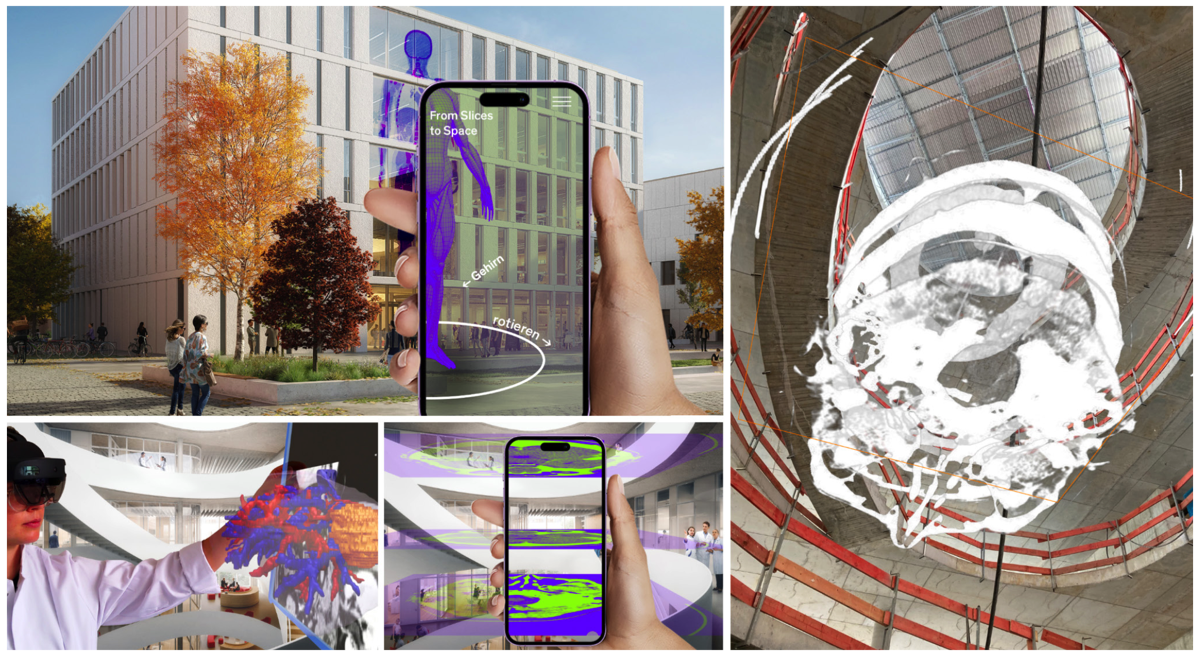
Under the title "From Slices to Spaces", the tandem of Moritz Queisner and Frédéric Eyl is implementing a science parcours in which spatially complex research data from surgery and biomedicine will be made multisensually accessible to a broad audience through new visualization techniques. Building on research work on new imaging techniques by Moritz Queisner, they employ Extended Reality techniques. Due to their unique ability to link digital objects with the real environment of the viewers, the 4D images they generate are particularly suited for representing and conveying spatial information.

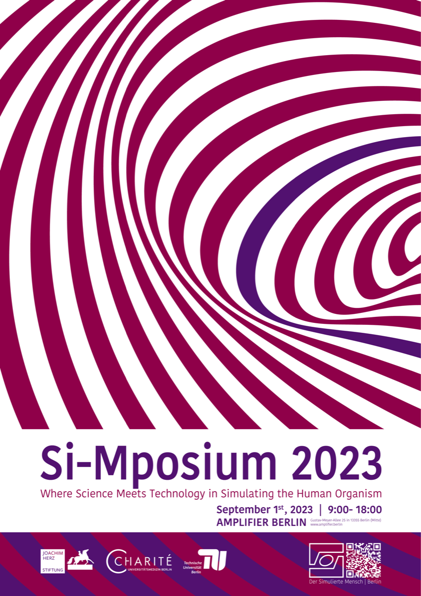
This is where the tandem's project comes in: 4D images are not only interesting for researchers to understand complex research data but can also provide laypeople with a less presupposing insight into research data and processes. Frédéric Eyl's media expertise will be used to make the specific visual knowledge from research comprehensible and experiential for non-experts. The science parcours is intended to integrate as a digital extension into the architecture of the new research building, "Der Simulierte Mensch", located on the premises of Charité. The parcours will include the facade, the inter-floor airspace, and the central glass surfaces within the building as its stations. By enabling users to explore 4D research data within the architecture and investigate it using their own smartphones in an AR application, concrete practices and deployment locations of new image-based technologies become experiential and comprehensible. This project not only enhances the perception of Charité and the scientific location of Berlin but also opens up places of knowledge creation to the public, making practices and techniques of life sciences more visible.


Stacks Image 28388
Eriselda Keshi and Simon Moosburner CSP fellows
Stacks Image 28393
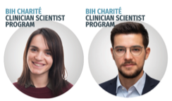
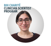
Dr. Eriselda Keshi and Dr. Simon Moosburner successfully applied for the BIH Charité Clinician Scientist Program (CSP).
The program provides a unique opportunity for young medical doc- tors to combine their clinical training with protected time for research. This structured career path fosters translation of scientific discoveries into application and strengthens the innovative capacity of academic medicine.
Participants of the CSP devote 50 percent of their working hours to research over a period of three years.

Dr. Keshi will work on "NeoPancreasPrint, a 3D printed islet hosting tissue based on biocompatible ink derived from human decellularized pancreas".
Dr. Moosburner applied with this project "Alleviation of Senescence induced Ischemia-Reperfusion in Liver Grafts of Elderly Donors [SenEx".


Congratulations!
Prof. Dr. Nathanael Raschzok
Stacks Image 28402
Nathanael Raschzok | Experimental Surgery | 2007

With us in the team since he was a student, he has so far climbed all academic levels with flying colours.
In recognition of his outstanding achievements in research, teaching and the promotion of young academics, Nathanael was awarded the title of Associate Professor at the Charité – Universitätsmedizin Berlin.

Congratulations, Prof. Raschzok!
DFG | Grant for Machine Perfusion RCT
Stacks Image 28414
The German Research Foundation (Deutsche Forschungsgemeinschaft – DFG) s sponsoring a multicenter randomized controlled clinical trial by Prof. Dr. Georg Lurje entitled "End-ischemic hypothermic oxygenated (HOPE) or normothermic machine perfusion (NMP) compared to conventional cold storage (CCS) in donation after brain death (DBD) liver transplantation; a prospective multicenter randomized controlled trial (HOPE-NMP)".

The purpose of this study is to test the effects of end-ischemic NMP versus end-ischemic HOPE technique in a multicentre prospective randomized controlled clinical trial (RCT) on ECD liver grafts in DBD liver-transplantation (HOPE-NMP). Two-hundred-thirteen (n = 213) human whole organ liver grafts will be submitted to either NMP (n = 85) or HOPE (n = 85) directly before implantation and going to be compared to a control-group of patients (n = 43) transplanted with static cold storage preserved ECD-allografts. Primary (surgical complications as assessed by the comprehensive complication index [CCI]) and secondary (graft- and patient survival, hospital costs, hospital stay) endpoints are going to be analysed.


Congratulations !
Work with us | PhD position
We offer a funded PhD position for a computational biologist @ Experimental Surgery, Charité – Universitätsmedizin Berlin!

Are you interested to work on cutting-edge cancer research, and investigating complex-chromosomal as well as other genomic phenomena in human carcinoma cells ?

  • You will work on complex genome analyses (single-cell analyses, whole genome analyses) to detect, annotate and re-construct circular DNA using state of the art computational tools (e.g., Amplicon architect).
  • We provide motivation and high commitment in supervision in areas around experimental oncology and surgery.
  • The position allows the applicant to pursue an academic qualification, while collaborating and networking with international experts on extrachromosomal circular DNA and pancreatic carcinogenesis.

Apply now!
Send brief cover letter and CV via email to Dr. med. Matthäus Felsenstein (matthaeus.felsenstein@charite.de)
<<  Page 2 / 12  >>
© 2025 Prof. Dr. Igor M. Sauer | Charité - Universitätsmedizin Berlin | Disclaimer

This website or its third-party tools use cookies, which are necessary to its functioning and required to achieve the purpose illustrated in the Disclaimer. By closing this banner, scrolling this page, clicking a link or continuing to browse otherwise, you agree to the use of cookies.